more on forensics - Page 2

Created

Last reply

Replies

22

Views

2.3k

Users

7

Frequent Posters

arni thumbnail
Posted: 19 years ago
#11
hi
i am trying doing it again plz do tell if it works!
arni thumbnail
Posted: 19 years ago
#12

DNA Examinations

Blood Saliva and Urine
Semen Hair
Buccal (oral) Tissues, Bones, and Teeth


Deoxyribonucleic acid (DNA) is analyzed in body fluids, stains, and other biological tissues recovered from evidence. The results of DNA analysis of questioned biological samples are compared with the results of DNA analysis of known samples. This analysis can associate victim(s) and/or suspect(s) with each other or with a crime scene.

There are two sources of DNA used in forensic analyses. Nuclear DNA (nDNA) is typically analyzed in evidence containing blood, semen, saliva, body tissues, and hairs that have tissue at their root ends. Mitochondrial DNA (mtDNA) is typically analyzed in evidence containing naturally shed hairs, hair fragments, bones, and teeth.

Questions concerning nuclear DNA evidence should be directed to 703-632-8446. Questions concerning mitochondrial DNA evidence should be directed to 703-632-7572. Follow the Evidence Submission directions including Requesting Evidence Examinations and Packaging and Shipping Evidence.

Documenting, Collecting, Packaging, and Preserving DNA Evidence

If DNA evidence is not properly documented, collected, packaged, and preserved, it will not meet the legal and scientific requirements for admissibility in a court of law.

    If DNA evidence is not properly documented, its origin can be questioned.

  • If it is not properly collected, biological activity can be lost.
    If it is not properly packaged, contamination can occur.

  • If it is not properly preserved, decomposition and deterioration can occur.

When DNA evidence is transferred by direct or secondary (indirect) means, it remains on surfaces by absorption or adherence. In general, liquid biological evidence is absorbed into surfaces, and solid biological evidence adheres to surfaces. Collecting, packaging, and preserving DNA evidence depends on the liquid or solid state and the condition of the evidence.

The more that evidence retains its original integrity until it reaches the Laboratory, the greater the possibility of conducting useful examinations. It may be necessary to use a variety of techniques to collect suspected body fluid evidence.

Blood Examinations
Examinations can determine the presence or absence of blood in stains. Examinations can also determine whether blood is human or not. Blood examinations cannot determine the age or the race of a person. Conventional serological techniques are not adequately informative to positively identify a person as the source of a stain.

Collecting Known Samples

Blood

    Only qualified medical personnel should collect blood samples from a person.

  • Collect at least two 5-mL tubes of blood in purple-top tubes with EDTA as an anticoagulant for DNA analysis. Collect drug- or alcohol-testing samples in gray-top tubes with NaF (sodium fluoride).
    Identify each tube with the date, time, subject's name, location, collector's name, case number, and evidence number.

  • Refrigerate, do not freeze blood samples. Use cold packs, not dry ice, during shipping.
    Pack liquid blood tubes individually in Styrofoam or cylindrical tubes with absorbent material surrounding the tubes.

    Label the outer container KEEP IN A COOL DRY PLACE, REFRIGERATE ON ARRIVAL, and BIOHAZARD.

  • Submit to the Laboratory as soon as possible.

Blood on a Person
    Absorb suspected liquid blood onto a clean cotton cloth or swab. Leave a portion of the cloth or swab unstained as a control. Air-dry the cloth or swab and pack in clean paper or an envelope with sealed corners. Do not use plastic containers.

  • Absorb suspected dried blood onto a clean cotton cloth or swab moistened with distilled water. Leave a portion of the cloth or swab unstained as a control. Air-dry the cloth or swab and pack in clean paper or an envelope with sealed corners. Do not use plastic containers.

Blood on Surfaces or in Snow or Water
    Absorb suspected liquid blood or blood clots onto a clean cotton cloth or swab. Leave a portion of the cloth or swab unstained as a control. Air-dry the cloth or swab and pack in clean paper or an envelope with sealed corners. Do not use plastic containers.

  • Collect suspected blood in snow or water immediately to avoid further dilution. Eliminate as much snow as possible. Place in a clean airtight container. Freeze the evidence and submit as soon as possible to the Laboratory.


Bloodstains

    Air-dry wet bloodstained garments. Wrap dried bloodstained garments in clean paper. Do not place wet or dried garments in plastic or airtight containers. Place all debris or residue from the garments in clean paper or an envelope with sealed corners.

    Air-dry small suspected wet bloodstained objects and submit the objects to the Laboratory. Preserve bloodstain patterns. Avoid creating additional stain patterns during drying and packaging. Pack to prevent stain removal by abrasive action during shipping. Pack in clean paper. Do not use plastic containers.

    When possible, cut a large sample of suspected bloodstains from immovable objects with a clean, sharp instrument. Collect an unstained control sample. Pack to prevent stain removal by abrasive action during shipping. Pack in clean paper. Do not use plastic containers.

  • Absorb suspected dried bloodstains on immovable objects onto a clean cotton cloth or swab moistened with distilled water. Leave a portion of the cloth or swab unstained as a control. Air-dry the cloth or swab and pack in clean paper or an envelope with sealed corners. Do not use plastic containers.


Blood Examination Request Letter

A blood examination request letter must contain the following information:

    A brief statement of facts relating to the case.

    Claims made by the suspect(s) regarding the source of the blood.

    Whether animal blood is present.

    Whether the stains were laundered or diluted with other body fluids.

  • Information regarding the victim(s)' and suspect(s)' health such as AIDS, hepatitis, or tuberculosis.

Semen and Semen Stains
    Absorb suspected liquid semen onto a clean cotton cloth or swab. Leave a portion of the cloth or swab unstained as a control. Air-dry the cloth or swab and pack in clean paper or an envelope with sealed corners. Do not use plastic containers.

    Submit small suspected dry semen-stained objects to the Laboratory. Pack to prevent stain removal by abrasive action during shipping. Pack in clean paper. Do not use plastic containers.

    When possible, cut a large sample of suspected semen stains from immovable objects with a clean, sharp instrument. Collect an unstained control sample. Pack to prevent stain removal by abrasive action during shipping. Pack in clean paper. Do not use plastic containers.

  • Absorb suspected dried semen stains on immovable objects onto a clean cotton cloth or swab moistened with distilled water. Leave a portion of the cloth or swab unstained as a control. Air-dry the swab or cloth and place in clean paper or an envelope with sealed corners. Do not use plastic containers.

Seminal Evidence From Sexual Assault Victim(s)
  • Sexual assault victim(s) must be medically examined in a hospital or a physician's office using a standard sexual assault evidence kit to collect vaginal, oral, and anal evidence.
  • Refrigerate and submit the evidence as soon as possible to the Laboratory.

Buccal (oral) swabs
    Use clean cotton swabs to collect buccal (oral) samples. Rub the inside surfaces of the cheeks thoroughly.

    Air-dry the swabs and place in clean paper or an envelope with sealed corners. Do not use plastic containers.

    Identify each sample with the date, time, subject's name, location, collector's name, case number, and evidence number.

  • Buccal samples do not need to be refrigerated.


Saliva and Urine

    Absorb suspected liquid saliva or urine onto a clean cotton cloth or swab. Leave a portion of the cloth unstained as a control. Air-dry the cloth or swab and pack in clean paper or an envelope with sealed corners. Do not use plastic containers.

    Submit suspected small, dry saliva- or urine-stained objects to the Laboratory. Pack to prevent stain removal by abrasive action during shipping. Pack in clean paper or an envelope with sealed corners. Do not use plastic containers.

  • When possible, cut a large sample of suspected saliva or urine stains from immovable objects with a clean, sharp instrument. Collect an unstained control sample. Pack to prevent stain removal by abrasive action during shipping. Pack in clean paper. Do not use plastic containers.
    Pick up cigarette butts with gloved hands or clean forceps. Do not submit ashes. Air-dry and place the cigarette butts from the same location (e.g., ashtray) in clean paper or an envelope with sealed corners. Do not submit the ashtray unless a latent print examination is requested. Package the ashtray separately. Do not use plastic containers.

    Pick up chewing gum with gloved hands or clean forceps. Air-dry and place in clean paper or an envelope with sealed corners. Do not use plastic containers.

  • Pick up envelopes and stamps with gloved hands or clean forceps and place in a clean envelope. Do not use plastic containers.

Hair
  • Pick up hair carefully with clean forceps to prevent damaging the root tissue.
  • Air-dry hair mixed with suspected body fluids.
    Package each group of hair separately in clean paper or an envelope with sealed corners. Do not use plastic containers.

  • Refrigerate and submit as soon as possible to the Laboratory.
Tissues, Bones, and Teeth

Call the Laboratory at 703-632-7572 prior to submitting suspected tissues, bones, or teeth to ensure that the evidence will be accepted for examination.
The communication accompanying the evidence must reference the telephone conversation accepting the evidence.

    Pick up suspected tissues, bones, and teeth with gloved hands or clean forceps.

  • Collect 1-2 cubic inches of red skeletal muscle.
  • Collect 3-5 inches of long bone such as the fibula or femur.
    Collect teeth in the following order:
      nonrestored molar.

      nonrestored premolar.

      nonrestored canine.

      nonrestored front tooth.

      restored molar.

      restored premolar.

    1. restored canine.

    2. restored front tooth.
  • Place tissue samples in a clean, airtight plastic container without formalin or formaldehyde. Place teeth and bone samples in clean paper or an envelope with sealed corners.
  • Freeze the evidence, place in Styrofoam containers, and ship overnight on dry ice.
< name=forsbar> </> Crime Scene Processing.
Much of what has been discussed previously is involved in crime scene processing, so there is little need to rehash. There remain a number of points that fit into the categories of trace evidence, the homicide victim and a few administrative matters to get the nitty gritty explanation of forensic science out the way and conclude this set of web pages. This is more the loose ends section than anything, a smattering of quick explanations for topics that don't appear in the other categories. But first, some review. Most physical evidence concerns itself with class characteristics and individual characteristics. Class characteristics are those characteristics which are common to a group of similar objects. For example, you buy a pair of Air Jordan sneakers. All Air Jordan sneakers have the same shape and same tread design on the bottom. These are class characteristics. Individual characteristics are those characteristics which are unique to a given object and set it apart from similar objects. You wear your Air Jordans around for awhile and they get worn. The treads wear down. They get little pits and gouges in them. These little pits and gouges are individual to your shoes and no others since no one has walked over the exact same surfaces in the exact same way in their Air Jordans. These concepts apply to all kinds of forensic evidence, from soil to glass, from rope to hefty bags. The fracture match is another important concept, particularly when trace evidence is concerned. Tear a piece of paper in half. Hold the two halves together. This is called a fracture match. No two tears are exactly alike. One half of a tear can always be matched back to its other half. Remember that. If a half of something found at a crime scene can be matched to the other half of something found on a suspect, that's damn good physical evidence. Crime Scene Documentation. The first thing you do after securing a crime scene is document it. Always take pictures. They are the best record available. They show the crime scene as it was found; where objects are in relation to other objects, victims, rooms, etc. Take notes. Describe the scene, its over all conditions. Describe rooms, lights, shades, locks, food; anything that can indicate a time frame, condition of scene or that might have even the slightest evidentiary significance. Check dates on mail and newspapers. Diagram the crime scene. Take measurements. Photos are good to show where an object is in relation to another object, but measurements tell exactly how far. Chain of Custody. Chain of Custody is of paramount importance to any investigation. It is the unbroken sequence of events that is caused by an item of evidence from the time it is found at the crime scene to the time it appears in court. Every link in this chain is documented, from discovery at the crime scene, through evidence gathering, storage, lab analysis, return to storage, transfer to court. Every link is documented by date, time, handling individual, what was done with the evidence by that individual. If chain of custody is broken, if the evidence cannot be accounted in one step of its journey from crime scene to court room, it is rendered inadmissible; useless to the case. Locard's Exchange Principal. Every time an individual comes in contact with a place or another individual, something of that individual is left behind at the place, and something of that place is taken away with the individual. If your Aunt Bertha gives you a big hug and walks away, fibers from her clothes will be on your clothes and fibers from your clothes will be on hers. Your hair is constantly falling out (circle of life, guys). You leave it all over the place. Just look around your house. You pick up carpet fibers on your shoes, dirt from the ground. Your skin flakes off. Look at the Wayne Williams case in Atlanta. He was convicted because fibers found on the body of one of his victims matched fibers from the carpet in his house. Might not sound like much, but it's GREAT physical evidence. Entomology. To paraphrase Indiana Jones, "Bugs. I hate bugs..." But they are great evidence and can be used to determine time of death. In some cases, they can be used to determine if a body has been moved from one geographic location to another. Certain bugs incubate and hatch at certain known rates. If bugs are found on a corpse, the age of the bugs can be extrapolated backward to estimate time of death. Blood Spatters. Blood spatters help a great deal in recontructing a crime scene. They can be used to corroborate or disprove and alibi. They can be used to convict the guilty. There is much more to it than looking at a stain or spatter and saying, "This is where the crime took place." The patterns of the spatters and the shapes of the individual blood droplets themselves can tell how the crime was committed. Drops falling from different heights (i.e. at different speeds) will leave different looking spatters. A drop falling from a low height of a few inches will leave a small cohesive circle. At greater heights, the circle will be larger and may even have a 'crown' effect. Hitting a surface at an angle does even more to disrupt a blood droplet. Perpendicular impact leaves a droplet fairly uniform, as shown below. A droplet hitting a surface at an angle will bulge out in one direction, indicating the direction of travel of the droplet. Cast off stains are a result of blunt force trauma (beating with an object such as a hammer). Pulling back from a blow produces a blood spatter that indicates direction, by creating an arc of blood droplets. You can determine the number of blows inflicted by counting the arcs. You can also determine the orientation of the individuals involved, the size of the object used and the right or left handedness of the assailant. Click here to go to the next page.
Glass Examinations
Glass comparison examinations can determine whether particles of glass originated from a broken source of glass. Glass fracture examinations can determine the direction and type of the breaking force and the sequencing of shots.

Questions concerning glass evidence should be directed to 703-632-7690. Follow the Evidence Submission directions including Requesting Evidence Examinations and Packaging and Shipping Evidence.

Comparison
  • Submit samples of glass from each broken window or source in leakproof containers such as film canisters or plastic pill bottles. Do not use paper or glass containers.
    Submit samples of laminated glass (e.g., windshield) from each side of the glass. Label the samples INSIDE and OUTSIDE and package separately in leakproof containers such as film canisters or plastic pill bottles. Do not use paper or glass containers.

    Submit the victim(s)' and suspect(s)' air-dried clothing. Each item must be packaged separately in a paper bag.

    Search for particles in the victim(s)' and suspect(s)' hair, skin, and wounds. Submit particles in leakproof containers such as film canisters or plastic pill bottles. Do not use paper or glass containers.

    Search for particles in vehicles by vacuuming each section of the vehicle separately. Do not use tape for recovering glass particles. Submit vacuum sweepings in leakproof containers. Do not use paper or glass containers.

    Ship known and questioned debris separately to avoid contamination.

  • Do not process evidence for latent prints.
Fracture
    Label the sides of the glass in the frame (INSIDE and OUTSIDE). Label the glass where it was removed in the frame (TOP, BOTTOM, LEFT, and RIGHT).

    Submit all glass pieces so that the pieces can be fitted together to identify the radial cracks near and at the point(s) of impact and to increase the probability of matching edges. Pack all glass separately and securely to avoid shifting and breaking during shipping.

    Submit the entire piece of laminated glass, if possible. Secure the glass between plywood or sturdy cardboard. Do not place any objects into the impact area.

  • Do not process evidence for latent prints.
< name=forsbar> </> Forensic Photography.
I'm not going to go into the basics of photography here. There are other pages on the web that will do it much better than I can. Actually, this section is going to be pretty short. There are just a few particulars to hit on, and a couple of interesting techniques. The first thing that needs to be done after securing the crime scene is photographing it. This creates a permanent record of the condition of the crime scene, one that is incontestable. First, take a picture that shows where the scene is; a shot with a street sign with the crime scene location in the background. Take pictures of the areas around the crime scene; alleys, dumpsters, rear areas, neighboring structures and even the structures across the street. Next, take pictures of the outside of the structure, showing points of entry and exit. Enter the structure, taking shots that show the locations and layout of the rooms. Take pictures of the whole room where the crime took place. Take close-ups of the scene or body. All pictures of items of evidence, which will be covered in the next paragraph, should be take both with and without a scale (a small ruler showing the size of the object). Take pictures with the scale to show the size of an object. Take pictures without the scale in case its presense in the picture blocks other evidence. What items are photographed at a crime scene? Bullet casings; photograph as a group and photograph individually. Photograph any dropped items, foot prints or animal tracks. If a homicide, photograph the body or bodies. Photograph any toolmarks, bitemarks or skin impressions. Basically, anything that might be evidence is photographed. Imprint evidence requires extra measures. Shoe imprints are photographed individually and as a series or group. Shoe imprints need to be lit from the side to show as much detail in the imprint as possible. Tire imprints are photographed from above as a whole. If the tire imprint is four feet long, then a picture showing all four feet is taken. Detail pictures are then taken showing one foot sections, each picture overlapping the one before it. This way, specific detail can be show and the overlapping pictures lined up to show the whole print. Again, all pictures are take with and without a scale. There is a special technique for no light situations. This technique is useful outdoors at night (perhaps a car accident scene), or in situations where the room is too big to light or there is no light available for pictures to be taken (such as a burnt out warehouse arson). The camera is set on a tripod with its shutter locked open. The photographer walks to several points in the room, popping off the flash, which is held in his or her hand. Each time the flash goes off, the film in the camera is exposed to another part of the room. The photographer does not appear as he/she is behind the flash and does not get exposed to the light when it pops off and only moves around the room while it is still dark. Remember, the film in a camera captures light. If there is no light, you can walk around in front of a camera all you want and never show up on the film. Video is also used to film crime scenes, taking long sweeping shots that take in everything in an unbroken time frame. The problem with video is, camcorder microphones will pick up the officers talking in the background, which can sometimes be embarrassing when the tape is replayed in court.
Gunshot Residue on Hands Examinations

For muzzle-to-target distance determinations involving gunshot residue, see Firearms Examinations.

When a firearm is discharged, vaporous and particulate materials called gunshot residue (GSR) are expelled. After collecting gunshot residue from a suspected shooter's hands, the major elemental components of most cartridge primer mixtures can be analyzed to associate a suspect with the recent discharge of gunpowder from a firearm. This examination is used to determine if a person was in the presence of gunshot residue within a limited time period after a weapon discharge.

The Laboratory provides gunshot residue examinations to assist FBI field office investigations only.

Questions concerning gunshot residue evidence should be directed to 703-632-8441. Follow the Evidence Submission directions including Requesting Evidence Examinations and Packaging and Shipping Evidence.
    Only gunshot residue evidence obtained using a collection kit designed for scanning electron microscopy will be accepted. Call the Laboratory at 703-632-8441 regarding the appropriate collection kits.

    Usually gunshot residue examinations will only be performed when samples are collected from living persons' hands.

    Gunshot residue evidence must be collected within five hours of exposure to the discharge of a firearm.

    Complete the information sheet included with the gunshot residue collection kit, and follow the instructions for collecting evidence.

  • Collecting gunshot residue samples requires five adhesive lifts suitable for scanning electron microscopic analysis. Dab the adhesive side of the stub against the surface (right palm, back of right hand, left palm, back of left hand). Use one stub per sampling surface. The remaining stub will be used as a control. Label each sampling surface stub (e.g., RIGHT PALM, BACK OF RIGHT HAND). Cap and seal the stubs in separate, resealable plastic bags.

Hairs and Fibers Examinations

Hairs
Hair examinations can determine whether hairs are animal or human. Race, body area, method of removal, damage, and alteration (e.g., bleaching or dyeing) can be determined from human hair analysis. Examinations can associate a hair to a person on the basis of microscopic characteristics in the hair but cannot provide absolute personal identification. Hairs that are associated will be submitted for mitochondrial DNA analysis. The animal species and family can be determined from hair analysis.

Fibers
Fiber examinations can identify the type of fiber such as animal (wool), vegetable (cotton), mineral (glass), and synthetic (manufactured). Questioned fibers can be compared to fibers from victim(s)' and suspect(s)' clothing, carpeting, and other textiles. A questioned piece of fabric can be physically matched to known fabric. Fabric composition, construction, and color can be compared, and impressions on fabric and from fabric can be examined. Clothing manufacturers' information can be determined by label searches.

Questions concerning hairs and fibers evidence should be directed to 703-632-8449. Follow the Evidence Submission directions including Requesting Evidence Examinations and Packaging and Shipping Evidence.

    Collect at least 25 known hairs from different parts of the head and/or pubic region. Comb and pull out the hairs. Submit hairs in clean paper or an envelope with sealed corners.

  • When possible, submit the entire garment or textile. Submit fibers in clean paper or an envelope with sealed corners.
Image Analysis Examinations

Photographic Comparisons
Examinations of film, negatives, digital images, photographic prints, and video recordings including surveillance images involve comparisons of subject(s) depicted in the questioned images with known images (e.g., photographs, videos) of suspect(s). Similar comparisons can be made between the subject(s)' clothing and clothing seized from the suspect(s). Comparisons can also be made with firearms, vehicles, and other objects depicted in surveillance images.
Photogrammetry
Dimensions can be derived from photographic images through the use of geometric formulae or on-site comparison. Examples of photogrammetry include determining the height of bank robbery subject(s) and the length of the weapon(s) used by the subject(s) depicted in the surveillance films.
Location, Time, and Date
Examinations of photographic evidence can determine the location, time, and date that an image was taken.
Authenticity and Image Manipulation Detection
Photographic evidence including film, video, and digital images can be examined to determine whether the image is the result of a composite, an alteration, or a copy.
Source and Age
Photographic products including film and prints can be dated, and the source can be established by examining manufacturing characteristics. This can establish the time frame during which a photograph was taken.
Cameras
Cameras seized as evidence can be examined to determine whether a specific camera exposed a specific image. Digital cameras, including digital-video cameras, can be compared with digital images and video clips to determine whether a specific camera captured a specific image or video clip.
Videos
Black-and-white and color photographic images can be produced from video images for enlargement and used in courtroom presentations.
Automobile Make and Model Identification
Vehicles depicted in surveillance images can be compared with the National Automotive Image File to determine make and model.
Child po*nography Examinations
The seized images of child po*nography can be compared against images in the Child Exploitation and Obscenity Reference File to identify the source of the images. Video clips can be examined to determine if any of the people and scenes depicted in the video clips are also recorded as still images in the File. Video clips and still images can also be examined to determine if they depict recordings or images of real people and events or whether they represent computer-generated subjects and events.

Questions concerning image analysis examinations should be directed to 703-632-6222. Questions concerning image analysis evidence should be directed to 703-632-6191. Follow the Evidence Submission directions including Requesting Evidence Examinations and Packaging and Shipping Evidence.

    Write-protect the original recording. Never use the Pause operation when viewing original video recordings.

    Submit original evidence (e.g., film or videotape) whenever possible because it contains the greatest level of detail. If the originals are unavailable, submit first generation photographic prints or videotapes.

    Process all film, including bank surveillance film, prior to submitting.

    When requesting forensic examinations based on video images, queue the original videotape to the approximate time of the pertinent area. State in a communication the date and time of the pertinent area and use the date-time stamp on the images or the counter indicator (set from the beginning of the tape at 000). If prints from the relevant frames are available, submit them for reference.

    Arrest or known photographs of suspect(s) for comparison with questioned images must depict the suspect(s) from many angles similar to the questioned images. If a facial comparison is requested, ensure that the suspect(s)' face or head fills more than half of the frame. If questioned images show tattoos or marks, include photographs of the same areas on the known suspect(s)' body.

    When taking known photographs for comparison with questioned images, use 35mm black-and-white film. If color film is used, include a color chart in the photographs.

    Do not mark or cut items submitted for comparison (e.g., clothing or firearms) where they are visible in the questioned images.

    Physical items such as clothing and firearms must first be submitted to the Laboratory for other examinations before they are submitted for image comparison.

    If photogrammetry is requested, include the dimensions of the scene to the nearest 1/8 inch and include a diagram or print from the surveillance film indicating the location of the measurements. Include one diagram or print for every angle used in the scene. Do not touch or move surveillance cameras except to remove the film.

    Submissions for comparison with the Child Exploitation and Obscenity Reference File must be limited to no more than 30 images. Call 703-643-6222 for specific instructions.

  • Address the outer container as follows:

    FEDERAL BUREAU OF INVESTIGATION
    ENGINEERING RESEARCH FACILITY
    ATTENTION: FORENSIC PROGRAM
    BUILDING 27958A
    QUANTICO VA 22135

Ink Examinations
Examining inked writing in conjunction with other techniques (e.g., handwriting analysis, watermark identification) can provide details regarding document preparation. The composition of writing inks varies with the type of writing instrument (e.g., ballpoint pen, fountain pen, porous-tip pen) and the date of the ink manufacture. In general, inks are composed of dyes in solvents and other materials that impart selected characteristics. Ink analysis is usually limited to comparisons of the organic dye components. When ink formulations are the same, it is not possible to determine whether the ink originated from the same source to the exclusion of others. Examinations cannot determine how long ink has been on a document.

Questions concerning ink evidence should be directed to 703-632-8441. Follow the Evidence Submission directions including Requesting Evidence Examinations and Packaging and Shipping Evidence.

  • Pack ink evidence separately from any document or surface with ink marks.
< name=forsbar> </> Forensic Photography.
I'm not going to go into the basics of photography here. There are other pages on the web that will do it much better than I can. Actually, this section is going to be pretty short. There are just a few particulars to hit on, and a couple of interesting techniques. The first thing that needs to be done after securing the crime scene is photographing it. This creates a permanent record of the condition of the crime scene, one that is incontestable. First, take a picture that shows where the scene is; a shot with a street sign with the crime scene location in the background. Take pictures of the areas around the crime scene; alleys, dumpsters, rear areas, neighboring structures and even the structures across the street. Next, take pictures of the outside of the structure, showing points of entry and exit. Enter the structure, taking shots that show the locations and layout of the rooms. Take pictures of the whole room where the crime took place. Take close-ups of the scene or body. All pictures of items of evidence, which will be covered in the next paragraph, should be take both with and without a scale (a small ruler showing the size of the object). Take pictures with the scale to show the size of an object. Take pictures without the scale in case its presense in the picture blocks other evidence. What items are photographed at a crime scene? Bullet casings; photograph as a group and photograph individually. Photograph any dropped items, foot prints or animal tracks. If a homicide, photograph the body or bodies. Photograph any toolmarks, bitemarks or skin impressions. Basically, anything that might be evidence is photographed. Imprint evidence requires extra measures. Shoe imprints are photographed individually and as a series or group. Shoe imprints need to be lit from the side to show as much detail in the imprint as possible. Tire imprints are photographed from above as a whole. If the tire imprint is four feet long, then a picture showing all four feet is taken. Detail pictures are then taken showing one foot sections, each picture overlapping the one before it. This way, specific detail can be show and the overlapping pictures lined up to show the whole print. Again, all pictures are take with and without a scale. There is a special technique for no light situations. This technique is useful outdoors at night (perhaps a car accident scene), or in situations where the room is too big to light or there is no light available for pictures to be taken (such as a burnt out warehouse arson). The camera is set on a tripod with its shutter locked open. The photographer walks to several points in the room, popping off the flash, which is held in his or her hand. Each time the flash goes off, the film in the camera is exposed to another part of the room. The photographer does not appear as he/she is behind the flash and does not get exposed to the light when it pops off and only moves around the room while it is still dark. Remember, the film in a camera captures light. If there is no light, you can walk around in front of a camera all you want and never show up on the film. Video is also used to film crime scenes, taking long sweeping shots that take in everything in an unbroken time frame. The problem with video is, camcorder microphones will pick up the officers talking in the background, which can sometimes be embarrassing when the tape is replayed in court.
arni thumbnail
Posted: 19 years ago
#13

FINGERPRINTS.TK
The first on line site that will tell you all about "Fingerprints"


Patterns

Author
Chatroom
(new)
Comparisons
Contacts
Country rules
(new)
Discussion Group
Do it yourself
Incipient Ridges
F.A.Q.
Fingerprints
Links
Methods
Minutiae
New(s)

No Fingerprints
Patterns
Patterns %
Pores
Printing persons
Prints on Animals
School Project

Service
Strange Prints

Startpage

A brief tour in the world of fingerprints

Hello dear visitor,
Thank you for visiting my page, I presume that you want to now a little bit more about fingerprints.
On this page I will try to tell you what are fingerprints and what we can do with them.

History of fingerprints.
A very long time ago, about 1750 years before the birth of Christ, people in Babylon used fingerprints to sign their identity on clay tablets. The first time ink prints were used was in China around the year 220. And then fingerprints became a forgotten item.......
The first time someone wrote about it again was in 1686 a man called Malpighius. He described friction ridges in detail. In the year 1823 J.E. Purkynje discovered that fingerprints could be classified. And then slowly the use of fingerprints started:
1858 - Sir William Herschel in India demanded laborers to sign contracts with fingerprints.
1877 - Sir William Herschel advised to take fingerprints from prisoners.
1880 - Dr. Henry Faulds published in "Nature" in England for the use of fingerprints in crime scenes.
1882 - Gilbert Thompson used thumbprints in the USA on cheques to avoid fraud.
1892 - Sir Francis Galton published that fingerprints are unique and invariable in his book Finger
Prints. This was the basic for our modern fingerprinting science.
1897 - Sir Edward Henry introduced the famous Galton-Henry classification system.
1901 - 1910 A lot of countries started using fingerprints.
1924 - In America the Identification Division from the FBI started.

ALL ABOUT FINGERPRINTS:

Fingerprints are made of ridges on the upperskin on hands and feet of all people and some animals. These ridges make lines of different size's and forms.
Everytime a line stops or splits it's called a typica. The amount of typica (with no difference) make's it an identification or not. The amount of typica is in many country's different. In Holland we require 10 to 12
characteristic points with no difference


Here is an example of a fingerprint


The patron of the line's looks like waves in the sand of a beach or stripe's on a zebra!

There are 2 rules where the science of fingerprints is based on:
1) They are "permanent" in that they are formed in the fetal stage, prior to birth, and remain the same throughout lifetime, barring disfiguration by scarring, until sometime after death when decomposition sets in.
That means that the prints do not change during a life time. That's not all true because fingerprints do change, but the changes can be explained. If not, they can't be identified. The changes can be made by: flexibility from the skin, growing, a dirty finger, scarring, a wound, or a disease of the skin.

2) They are "unique" in that no two fingerprints, or friction ridge area, made by different fingers or areas, are the same (or are identical in their ridge characteristic arrangement).

People always ask if identical twins have the same prints, the answer is NO.
They have completely different fingerprints, although they have the same DNA.
Some people think that it is because there is a influence of the environs that make the prints develop different.
There is never found a print that is the same, even not on the same person.
The databanks of fingerprints all around the world, are containing billions of unique prints!

Types of prints.
A print is not only made of a collection of lines but the all the lines together
make a special figure. And there are not so very much different figures as you would presume. There are about 7 different types as you can see down here:


- Arch: The lines are going like waves from one site to the other site.
- Tentarch: Like the Arch but with a rising stick in the middle.
- Loop: The lines coming from one site returning in the middle to the te same
side
- Double loop: Like the loop but with two loops inside, one standing one
hanging
- Pocked loop: Like the loop but with a small circle in the turning point
- Whorl: The lines are making circles
- Mixed figure: Composed with different figures

But in every type of print there are lots of differences, the number of lines, their shape or their size make every figure different.


Just a bug!

Transmitting prints to a object.
- The most common way is leaving fat from the finger on the object.
- It is also possible that Amino-acids from the finger leave their marks.
- A finger can make a "stamp" in a soft underground like putty.
- A mark can be made by a substance on the finger like blood or paint.

Making marks visible.
There are a lot of ways to make marks visible. In fact a lot of marks can be seen if you look good. Just put a finger on a window and take a good look at the window. With the right light you can see the mark. It can be seen because grease from the fingertip puts a "stamp" of the finger on the glass. It will not be there forever because it simply will dry out. But there always will be a small residua left, if the object is not cleaned.
Fresh fingerprints can be found and made visible with powders that will stick on the grease of the finger. It are very fine powders, mostly is used aluminum powder.

The underground on which the print is put on is very important for the way the prints must be made visible. When a print is put on a piece of paper it is possible to make it visible with a magnetic iron powder. Strictly speaking the powder is not magnetic but the lifting brush is. This can only be done on very fresh marks, not much older than a few hours. After that an other method must be used: Ninhydrin. It is a chemical that colors the Amino-acids that are left behind after contact with the skin.

In a lot of cases involving metal or plastic objects the cyano-method can be used. When a few drops of superglue, cyano-acrylate, are heated they vaporize. The smoke of the cyano will attach to the fingerprints leaving a clear white print. It must be done in a special space under special conditions.
Do never try this at home because it is very poisonous!

Saving the marks.
It is possible to save the marks, for further investigation, by making
photographs of it, storing it on rubber lifters or tape, keeping the original
underground or copying it.

Down here you can see how fingerprints on a bottle are made visible with a brush and aluminum powder. A black rubber lifter is installed over the prints. After removing the black lifter, a copy of the prints is made. A plastic sheet is placed over the prints so they would not be damaged. The prints are now usable for further investigation.


1. Brush it with aluminum powder
2. The marks became visible.
3. Put a black rubber lifter on it.
4. The marks are on the lifter.
5. Put a plastic sheet over it.
6. Ready to search!

Typica.
A fingerprint contains a lot of typica, unique lines. They are short or long, they can start, stop or split, they make figures, can be big, small or broad.
And above all it can happen at every place. So now you can imagine that no two fingerprints are the same. It is a little bit like a living-room, in all the world there will be no two the same living-rooms. Flowers, paintings, pictures, trash, furniture and music will make every living-room unique!


Here you see a part of a fingerprint.

Generally there are two kinds of real typica:
- Ending lines
- Splitting lines (bifurcation's)
Above that there are a lot of other differences made up by those two typica: a very short line between two other lines is called an "isle", when a line splits and immediately returns to one line it is called an "eye". When it splits and one of the two splitting lines immediately returns to the site it came from and stops it will be called a "hook".
A lot of times one can see small lines or lineparts between two friction ridges. It looks a little like newborn lines, and in some rare cases it is seen that such a line became greater in a few years. But that are always very small parts of lines.

Identifications.
Worldwide there is no rule when a identification can be made.
In the Netherlands we require 12 characteristic points,
In South-Africa 7 points will make a identification,
And in a lot of countries (England, America) there is no rule, the expert decides.

If you want to see a fingerprint will all the typica click

Sign guestbook, suggestions or questions, press here!

Back to Top
< scrollAmount=9 scrollDelay=146 width=392 height=19 align="middle" border="0">copyright by H. v.d. Nieuwendijk, suggestions can be mailed to: ---> ---></> e--mail

FINGERPRINTS.TK
The first on line site that will tell you all about "Fingerprints"


Patterns

Author
Chatroom
(new)
Comparisons
Contacts
Country rules
(new)
Discussion Group
Do it yourself
Incipient Ridges
F.A.Q.
Fingerprints
Links
Methods
Minutiae
New(s)

No Fingerprints
Patterns
Patterns %
Pores
Printing persons
Prints on Animals
School Project

Service
Strange Prints

Startpage

School project, print out and ready!

mailto:dacty@xs4all.nl

1. What are fingerprints?


Everyone knows them, the grease fingers on a window. That are fingerprints we say. We do not think further.
But a fingerprint is more than a fat print on a piece of glass.

Most prints are not useful for a comparison, there is not enough information available in the print. Prints can be to old, to small, to dirty or damaged. Most fingerprints disappear after some days or some weeks. To know how to look at a print I am going to tell you the basics of fingerprints. .

The right definition of a fingerprint is strictly speaking the print (stamp) that a finger left on an object.

Besides fingerprints there are also other parts of the body that can leave good prints:

The inside of the fingerprint,
The inside of the hands,
The bottom of feet and toe.

To understand where we are talking about we have to take a good look at the tips of our fingers. Use a magnifying glass to see it better!

mailto:dacty@xs4all.nl

Her you see lines in the skin that are higher than the surface. The lines are going in a certain direction. They look like a small dike with a dry ditch in between. This lines we call friction ridges. Sometimes there are small islands in between. The friction ridges are not always the same length, and are not always going in the same direction.

Sometimes a line stops or splits.


That are the unique points.

mailto:dacty@xs4all.nl

And that is the clue for the great mystery of the fingerprint!

Because there are so many lines on a fingertip and because they are stopping, starting or slitting so many times there are no two persons in the world that have the same fingerprints. Even a square millimeter is different at all persons!

2. How does a finger leave a mark on an object?
First because there has been a contact between the finger and the object. The fingers can leave there marks on different ways on the object. It can be done on several ways:

The fat that is on the skin leaves a small layer on the object. That leaves a invisible stamp on the object. Sometimes you can see the stamp on a window.
The friction ridges (ditches) on the fingertip makes marks in a soft underground.
That happens in clay or putty.
The fingertip makes contact with a dusty or dirty underground. Than a part of the dirt will be removed because of that a negative print will arise. It can be seen on a dusty window-frame.
Acids in the fat of the fingertip etching in the surface of the object, For instance on copper.
A dirty finger can leave a mark, think of a finger with blood. The blood will leave a print.

3. Why are fingerprints important?


Searching with fingerprints has only one big reason:

To determinate to whom the print belongs.


An identification of an fingerprints is for 100% reliable. There is no other technique that can establish such a high standard.
Even DNA does not give absolute certainty. A twin does have the same DNA structure but does have totally different fingerprints.

mailto:dacty@xs4all.nl

The fact that everyone in the world has different fingerprints can be used in very different ways.
Her are some examples for the use of fingerprints:

Solving crimes,
Identity control,
Searching for identity with unknown dead,
Entrance control at important buildings,
Sometimes in a passport a fingerprint is included.

All around the world there are different standards to make an identification. In some countries it will be called an identification if there are somewhere between 7 to 15 identical points in the mark and fingertip. Other countries leave the identification over to the expert. It always is important that the are no unexplainable differences!

We talk about explainable differences for instance when there is a scar on the skin, dirt in the background or on the finger, warts or differences because of the flexibility of the fingertip.

4. How do we search with a mark?


To compare a mark it is necessary to have the prints of a person.

To take the fingerprints of a person most of the time ink is used. Some ink is put on the fingertip and than the fingertip is pushed on a piece of paper.
This can be done under force (crime suspects) but it also can be done in free will.

The fingerprinting is mostly done at the police. They take prints of fingertips and of the palm of the hand. In some cases prints can be made from the foot.
Then all those prints go to a central computer. That computer is used to search with the found marks.

To take someone's prints on free will can be done at persons that want to prove they are innocent or on identity papers. Sometimes prints are taken from unknown or dead people.

5. How can fingerprints made visible?


There are a lot of techniques that make it possible to make invisible prints visible. And to store them for further investigation.

The mostly used method is with powders. The powder will stick on the fat that has left there by the fingertip.
To store this mark it is possible to make a picture of it but an much easier way is done with a rubber foil. The foil is pushed over the mark and the print will be lifted on the foil. The print on the foil can be used for further investigation.

A method that is used very much is working with superglue. A little bit of superglue is being heated and it will vaporize. The vapor attaches to the fingerprint and that will become visible. Do not try this at home because it is toxic and it must be done in a laboratory.

To find prints on paper you can use a fine iron powder. The iron will stick on the fingerprint.
To find older prints on paper ninhydrin (a chemical) is used. After a while it will color the prints purple!

There are a lot of other methods with all kinds of light or chemicals.

6. The way fingerprints makes figures.
The friction ridges are going parallel to each other and together they make a figure.

Global there are 3 groundshapes:
- Friction ridges making an arch,
- Friction ridges making a whorl,
- Friction ridges making an loop.

From the shapes above a lot of other figures can be made but these are the most important.

7. The structure of a fingerprint.
Most fingerprints have a shape where there is a center visible. That center is called "the core", see top pearl. Another place is the place where a lot of lines come together "the delta", see bottom pearl.

http://www.gironet.nl/home/hansnwdk

In a fingerprint there can be a lot of lines. But there can be a great different in the amount of lines between one person or the other.

8. The future of the fingerprint.
Because fingerprints will always exist the always will be used. In the future there will be a lot of computers that can search with fingerprints.

And in the future there will be the need for a lot of people that can work with fingerprints. Because a computer can search for a matching print but it can never tell if it is a real identification. There is always the need for people that have to check the results of the computer.

Hans van den Nieuwendijk, http://www.fingerprints.demon.nl/ E-mail mailto:fingerprints@fingerprints.demon.nl

< scrollAmount=9 scrollDelay=146 width=392 height=19 align="middle" border="0">copyright by H. v.d. Nieuwendijk, suggestions can be mailed to: ---> ---></> e--mail
arni thumbnail
Posted: 19 years ago
#14
by the way just for ur gk i wud like to tell tht
the "wysiwyg "editor at the end of the post reply box gets its name from the following fact
"WHAT YOU SEE IS WHAT YOU GET"
shefali_goenka thumbnail
19th Anniversary Thumbnail Voyager Thumbnail Networker 1 Thumbnail
Posted: 19 years ago
#15
maine bhi kuch information di hai.hope aap logo ko acchi lage.woh maine agle post maine diya hai.
shefali_goenka thumbnail
19th Anniversary Thumbnail Voyager Thumbnail Networker 1 Thumbnail
Posted: 19 years ago
#16


------------------------------------------------------------ --------------------

DNA Testing: An Introduction For Non-Scientists
An Illustrated Explanation

by DONALD E. RILEY, Ph.D.
University of Washington
(Copyright 1997-2005)

Revised Edition Posted April 6, 2005


------------------------------------------------------------ --------------------

INTRODUCTION

The explanation of DNA testing that follows is intended as an introduction to the subject for those who may have limited backgrounds in biological science. While basically accurate, this explanation involves liberal use of illustration and, in some cases, over-simplification. Although intended to be informative, this is brief and incomplete explanation of a complex subject. The author suggests consulting the scientific literature for more rigorous details and alternative views.

DNA EXPLAINED IN EASY TERMS

DNA is material that governs inheritance of eye color, hair color, stature, bone density and many other human and animal traits. DNA is a long, but narrow string-like object. A one foot long string or strand of DNA is normally packed into a space roughly equal to a cube 1/millionth of an inch on a side. This is possible only because DNA is a very thin string.

Our body's cells each contain a complete sample of our DNA. One cell is roughly equal in size to the cube described in the previous paragraph. There are muscle cells, brain cells, liver cells, blood cells, sperm cells and others. Basically, every part of the body is made up of these tiny cells and each contains a sample or complement of DNA identical to that of every other cell within a given person. There are a few exceptions. For example, our red blood cells lack DNA. Blood itself can be typed because of the DNA contained in our white blood cells.

Not only does the human body rely on DNA but so do most living things including plants, animals and bacteria.

A strand of DNA is made up of tiny building-blocks. There are only four, different basic building-blocks. Scientists usually refer to these using four letters, A, T, G, and C. These four letters are short nicknames for more complicated building-block chemical names, but actually the letters (A,T, G and C) are used much more commonly than the chemical names so the latter will not be mentioned here. Another term for DNA's building blocks is the term, "bases." A, T, G and C are bases.

For example, to refer to a particular piece of DNA, we might write: AATTGCCTTTTAAAAA. This is a perfectly acceptable way of describing a piece of DNA. Someone with a machine called a DNA synthesizer could actually synthesize the same piece of DNA from the information AATTGCCTTTTAAAAA alone.

The sequence of bases (letters) can code for many properties of the body's cells. The cells can read this code. Some DNA sequences encode important information for the cell. Such DNA is called, not surprisingly, "coding DNA." Our cells also contain much DNA that doesn't encode anything that we know about. If the DNA doesn't encode anything, it is called non-coding DNA or sometimes, "junk DNA."[1]

The DNA code, or genetic code as it is called, is passed through the sperm and egg to the offspring. A single sperm cell contains about three billion bases consisting of A, T, G and C that follow each other in a well defined sequence along the strand of DNA. Each egg cell also contains three billion bases arranged in a well-defined sequence very similar, but not identical to the sperm.

Both coding and non-coding DNAs may vary from one individual to another. These DNA variations can be used to identify people or at least distinguish one person from another.

What is a Locus?

A locus (with a hard "c", LOW-KUS) is simply a location in the DNA. The plural of locus is, loci ( with a soft "c", pronounced LOW-S-EYE). Again, the DNA is a long string like object as illustrated below. A locus is simply a location in the DNA. Such locations, or loci, reside at specific places on chromosomes.

What is a Chromosome?

When a cell is getting ready to divide creating two daughter cells, it packs its DNA into bundles called chromosomes. Chromosomes are just bundles of DNA. For humans, there are consistently 23 pairs of chromosomes, each with a consistent size and shape. Chromosomes are numbered. Chromosome number 1 is the largest chromosome; chromosome number 2 a little smaller and so on. Among the 23 pairs of chromosomes there is a pair called the sex chromosomes. This is something of a misnomer, since there are many functions on the "sex" chromosomes that have nothing to do with sex. In females, the sex-chromosome pair consists of two similar size chromosomes called X chromosomes. Males have one X and one small Y chromosome.


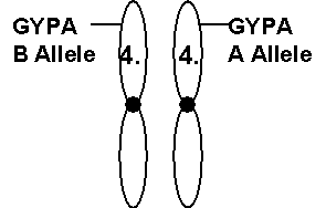
Unless it has been purified, our DNA is actually not a loosely tangled string as illustrated but rather is well organized and packaged into what are called chromosomes. A chromosome is a tightly folded bundle of DNA. Chromosomes are most visible when cells divide. In a microscope, chromosomes look something like this without the numbers and letters:


The illustration shows a pair of chromosomes named chromosome number 4, one pair among 23 pairs of chromosomes. The illustration also shows the position of a locus that happens to be called "GYPA." In this example, the chromosome on the left has the variation called the B allele while the chromosome on the right has the variation called the A allele.

What are alleles?

Alleles (ALL-EELS') are just variations at a particular site on a chromosome. Since each chromosome has a similar chromosome partner (except for males with their X and Y chromosomes) each locus is duplicated. Loci can vary a bit. If a person has two identical versions of the locus, they are said to be homozygous (HOMO-Z-EYE'-GUS). If there is a difference, they are said to be heterozygous (HETERO-Z-EYE'-GUS).


FORENSIC DNA TESTING

There have been two main types of forensic DNA testing. They are often called, RFLP and PCR based testing, although these terms are not very descriptive. Generally, RFLP testing requires larger amounts of DNA and the DNA must be undegraded. Crime-scene evidence that is old or that is present in small amounts is often unsuitable for RFLP testing. Warm moist conditions may accelerate DNA degradation rendering it unsuitable for RFLP in a relatively short period of time.

PCR-based testing often requires less DNA than RFLP testing and the DNA may be partially degraded, more so than is the case with RFLP. However, PCR still has sample size and degradation limitations that sometimes may be under-appreciated. PCR-based tests are also extremely sensitive to contaminating DNA at the crime scene and within the test laboratory. During PCR, contaminants may be amplified up to a billion times their original concentration. Contamination can influence PCR results, particularly in the absence of proper handling techniques and proper controls for contamination.

PCR is less direct and somewhat more prone to error than RFLP. However, PCR has tended to replace RFLP in forensic testing primarily because PCR based tests are faster and more sensitive.

RFLP EXPLAINED IN EASY TERMS

RFLP has been almost entirely replaced by PCR-based testing. The following description of RFLP is included here primarily for historic reasons (more current formats see below).

RFLP DNA testing has four basic steps:

1. The DNA from crime-scene evidence or from a reference sample is cut with something called a restriction enzyme. The restriction enzyme recognizes a particular short sequence such as AATT that occurs many times in a given cell's DNA. One enzyme commonly used is called Hae III (pronounced: Hay Three) but the choice of enzyme varies. For RFLP to work, the analyst needs thousands of cells. If thousands of cells are present from a single individual, they will all be cut in same place along their DNA by the enzyme because each cells DNA is identical to every other cell of that person.

2. The cut DNA pieces are now sorted according to size by a device called a gel. The DNA is placed at one end of a slab of gelatin and it is drawn through the gel by an electric current. The gel acts like a sieve allowing small DNA fragments to move more rapidly than larger ones.

3. After the gel has separated the DNA pieces according to size, a blot or replica of the gel is made to trap the DNA in the positions that they end up in, with small DNA fragments near one end of the blot and large ones near the other end. The blot is now treated with a piece of DNA called a probe. The probe is simply a piece of DNA that binds to the DNA on the blot in the position were a similar sequence (the target sequence) is located.

4. The size or sizes of the target DNA fragments recognized by the probe are measured. Using the same probe and enzyme, the test lab will perform these same steps for many people. These sizes and how they distribute among large groups of people form a database. From the database a rough idea of how common a given DNA size measured by a given probe is found. The commonness of a given size of DNA fragment is called a population frequency.


The restriction enzyme cuts the DNA into thousands of fragments of nearly all possible sizes. The sample is then electrophoretically separated. The DNA at this point is invisible in the gel unless the DNA is stained with a dye. A replica of the gel's DNA is made on something called a blot (also called a Southern blot) or membrane. The blot is then probed (mixed with) a special preparation of DNA that recognizes a specific DNA sequence or locus. Often, the probe is a radioactively labeled DNA sequence (represented by * labeled object in the figure above). Excess probe is washed off the blot, then the blot is laid onto X-ray film. Development reveals bands indicating the sizes of the alleles for the locus within each sample. The film is now called an "autorad." The band sizes are measured by comparing them with a "ladder" of known DNA sizes that is run next to the sample. A match may be declared if two samples have RFLP band sizes that are all within 5% of one another in size.

For RFLP analysis to be reliable, all complex steps of the analysis must be carefully controlled. Databases must be large meaning they include many people; they must be representative of the potential test subjects. Because of the complexities of populations, databases must be interpreted with extreme care. For example, DNA fragment sizes rare in one population may be very common in other populations. Further, sub-populations or populations within populations must be considered.

PCR EXPLAINED IN EASY TERMS

PCR is an abbreviation for "polymerase chain reaction." (POLL'-IM-ER-ACE). This term applies to a wide variety of different DNA tests that differ in reliability and effectiveness. Reliabilities of each kind of PCR test need independent verification. PCR itself doesn't accomplish DNA typing, it only increases the amount of DNA available for typing.

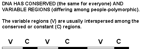
PCR uses constant regions of DNA sequence to prime the copying of variable regions of DNA sequence.


PCR typically uses two short pieces of known DNA called primers (small arrows below). These serve as starting points for the copying of a region of DNA.


Many forensic laboratories use commercial supplied DNA testing kits that contain key components for certain PCR-based tests. PM plus DQA1TM, Profiler PlusTM and CofilerTM and IdentifilerTM are all test kits commercially supplied by PE Applied Biosystems. PowerPlexTM is another test kit with variations supplied by Promega. PowerPlex kits have published primers, an advantage if the precise DNA targeted is to be recorded for posterity or studied for research. As of 2005, Profiler PlusTM and CofilerTM and PowerPlexTM are probably the most commonly used test kits in US forensic laboratories.

PCR Contamination

It is worth considering contamination early in this discussion since this is a well-recognized limitation. Unfortunately, the importance contamination in PCR is often underestimated. PCR copies DNA efficiently if the initial DNA is in good condition. A single DNA entity (molecule) can become millions or billions of DNA molecules in about three hours. The PCR process is sometimes compared to a Xerox machine since many copies are made. While initially, this is a useful comparison, it doesn't communicate the true, chain-reaction nature of PCR. In PCR, the original DNA is copied, then the copies are copied, those copies are copied and so on. This results in dramatic increases in the amount of DNA that couldn't be easily accomplished in the Xeroxing analogy. The PCR process deserves its classification as a "chain-reaction" because it has much in common with other chain reactions such as avalanches.

PCR is also very similar to what happens when a clinical infection occurs. Clinicians have known for many years that a single germ (bacterial cell or virus) contaminating a wound can produce a massive infection. Similarly, a DNA molecule can contaminate (infect) a PCR and become a significant problem. The ability of small amounts of DNA to produce false and misleading results is well-known and well-documented within the research community, where the technology originated. Anyone who has caught a cold from an unknown source, or who has a pollen allergy should have some sense of how easily PCRs are contaminated. Actually, it is probably easier to contaminate a PCR than to catch a cold since unlike our bodies, PCRs lack immune systems. The only protection PCRs have is the technique of the analyst, use of control samples to monitor contaminants and careful interpretation.

Prevention of false results involves the use of carefully applied controls and techniques. As described later, such controls and techniques can rarely guarantee that contamination hasn't influenced the results. In forensic DNA testing, some of the scientifically worst-case scenarios can be prevented by keeping DNA samples from known individuals well out of range of other items of evidence at all stages. Most forensic DNA laboratories perform negative controls, blank samples that will often detect contaminants in the laboratory. The blanks detect contaminants by showing partial or full DNA profiles representing the contaminants. Alternatively, the blank may show no profile, consistent with, but not proving that contamination didn't occur. Unfortunately, a few forensic DNA laboratories omit their controls. A few favor the controls by using special equipment on them, or by not carrying them through the entire procedure. Such practices are hazardous, especially when an important evidentiary sample has a low amount of DNA, degraded DNA, or otherwise presents as a minimal or partial (see below) sample. In short, while PCR is a useful research tool, all applications require extreme care and vigilance.

STRs
This will be presented in some detail because STRs are important in current, forensic DNA testing. The abbreviation, STR stands for Short Tandem Repeat. STRs are the type of DNA used in most of the currently popular forensic DNA tests. STR is a generic term that describes any short, repeating DNA sequence. For example, the DNA sequence ATATATATATAT is an STR that has a repeating motif consisting of two bases, A and T. It turns out that our DNA has a variety of STRs scattered among DNA sequences that encode cellular functions. For reasons that are not entirely understood, people vary from one another in the number of repeats they have, at least for some STR loci. For example, person #1 may have ATATAT at a particular locus while person #2 may have ATATATATATAT. Thus, STRs are often variable (polymorphic) and these variations are used to try and distinguish people. The term, STR doesn't necessarily imply PCR. PCR is one of many methods that might be used to help analyze STRs. STRs have also been analyzed by DNA sequencing for example. To understand PCR-assisted STR typing, it is useful to briefly consider how such PCRs are designed.

Suppose that laboratory data revealed the following DNA sequence:

--ATGCTAGTATTTGGATAGATAGATAGATAGATAGATAGATAAAAAAATTTTTTTT--

The STR is underlined and consists of the sequence, GATA repeated 7 times. The dashes at the beginning and end of the overall sequence shown indicate that there is more sequence available both upstream and downstream of the region shown. Remember, DNA is relatively very long and linear and we are just going to look at a small region of it.

Now, let's say we want to design a PCR to examine this same locus in other people. To design the PCR, we need two primers, short synthetic DNA molecules that recognize the region. One primer might be, ATGCTAGTA (Italics, in the above sequence) a sequence that would recognize the DNA flanking the left side of the STR. The second primer might be, AAAAAAAATTTTTT. This is called the downstream primer and it might be difficult to recognize in the sequence. The reason it is difficult to recognize at first is that it is the complement of the sequence, AAAAAAAATTTTTT (italics, on the right in the longer sequence above). See "General Considerations", for a more detailed discussion.

What is the complement of a DNA sequence? This might be more information than you would like, but to really understand PCR primers, try to walk through this:

The complement of a DNA sequence is the sequence written backwards exchanging all A's for T's, all T's for A's, all G's for C's and all C's for G's. For example, the complement of the sequence, AGTA is TACT. An easy way to get the complement of a DNA sequence is to write another line below the original sequence remembering that A replaces T and G replaces C. Then read the lower line backwards:

So, for the sequence:

GATCTTAGCTTTAAAGCCC

write the complementary line below it giving:

GATCTTAGCTTTAAAGCCC

CTAGAATCGAAATTTCGGG

Then, just read the lower line backwards (from right to left) giving the complement:

GGGCTTTAAAGCTAAGATC

In practical words, the upstream (left) primer can be a direct reading of the target sequence while the downstream primer (right) must be the complement of the directly read sequence.

If the above is confusing, it may suffice to think of the primers as two arrows that point at one another with the STR located between them. This is how the PCR targets the locus and the STR.

In practice, PCR primers are usually at least 17 bases in length. The point here is that to use PCR to target an STR, the primers recognize constant, conserved sequences that flank the actual STR. This means that the actual length of the target sequence depends on where the primers are placed in the flanking sequence. For example, the Promega and PE, Applied Biosystems test kits use mostly different primers. For example, the upstream primer could be designed to recognize DNA 100 bases upstream of the sequence shown. Similarly, the downstream primer could be designed to recognize DNA further downstream. Such placement of the primers by design, further upstream and downstream, would make all alleles (variations) of the STR appear to be larger than if the primers are placed by design close to the STR itself. Wherever the primers are placed, that defines the region we will examine. That region will then vary among individuals due to changes in the STR itself as explained above for the simple STR based on the repeating AT motif.


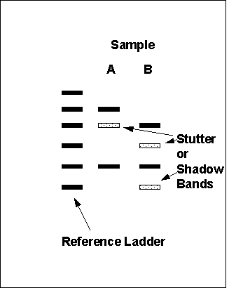
After PCR is used to provide many copies of a given person's STR, the products (copies) are separated according to size on an electrophoretic gel (see RFLP above for more details about gels). The gel can be flat, as for RFLP, or it can be in a round tube, called a capillary with a detector at the end of it. Typical flat gel STR results look like this:

The black bars are called bands. Each band is made up of many identical-size DNA molecules that were produced by PCR. The gel separates smaller bands (DNA molecules) from larger ones. The bands near the lower end of the gel are smaller (ie. the DNA fragments are shorter in length) than those near the top. For example, looking at the reference ladder, the first band near the lower end of the gel is the smallest STR. For simplicity, let's say this smallest band contains a single repeat such as CATG, flanked by other DNA that the primers actually recognize in everyone's DNA. The next higher band in the ladder would then contain 2 repeats, CATGCATG; the next 3 repeats and so on. By comparing the positions of bands in the unknown samples with the reference ladder, the allele sizes are deduced. In this example, Sample A had bands at the 2-repeat position and the 5-repeat position. Common terminology would call this sample a 2,5 type. Sample B would be called, 2,4. For a single person, each locus normally has two alleles and these can be different (heterozygous) or the same (homozygous).

Multiplex STR

One of the more commonly encountered STR test designs in forensic testing is called, Multiplex STR. There are multiplex, PCR reagent kits sold by both: PE Applied Biosystems and by Promega. Such systems combine three or more different PCRs in one reaction that targets distinct STR loci at the same time. Three of the commonly used loci are called, CSF1PO, TPOX and THO1. Again, the names of the loci have historical significance, but are of little importance as names.

Profiler Plus and CofilerTM (PE Applied Biosystems) combines 13 different STR loci. PowerplexTM (Promega) uses the same 13 loci but the primers used are different. The Promega kit incorporates published primer sequences, a significant scientific advantage, since without the primer sequences, it is unclear which STRs at some loci are targeted. A newer typing kit, IdentifilerTM (PE Applied Biosystems) incorporates the original 13 loci but adds 2 additional loci. By design (meaning where the primers were placed on the DNA by the designers) multiplex STR loci have different, non-overlapping size ranges so that DNA fragments from the different loci will have different, non-overlapping size ranges. Or, if the sizes overlap, they are tagged with differing dyes to help distinguish the 13 loci. These test systems have boldly ambitious designs and should be considered fairly experimental, especially for samples whose quantity and/or quality is outside tested limits.

There have been some discrepancies in profiles obtained with test kits from the two manufactures when the same samples were analyzed. These discrepancies are not extremely common but are noticeable and fairly dramatic when they occur. Any base within DNA can mutate (ie. change). For example, an A base at a particular position can change to a G. Such mutations usually first appear in a sperm or egg cell. Each mutation then appears throughout the body of the person who results from such a sperm or egg. Discrepancies in test kit results are thought to be due to mutations in the sites that the primers bind. These events are called, primer binding site mutations or PBS mutations.

Multiplex STRs are often combined with PCR for another locus called, amelogenin (pronunciation varies, but usually it is AM'-EEL-O-GEN-IN). Amelogenin adds little to the discriminating power of the test. Its purpose is to help distinguish male and female sources of DNA by detecting the X and Y chromosomes. The amelogenin products have sizes that place them outside the size ranges of the other loci.

Compared to PCR-based systems originally introduced, such as PM plus DQA1 (PE Applied Biosystems) multiplex STRs are technically more simple and direct at the allele detection stage. On the other hand, multiplex STR are slightly more vulnerable to missing alleles. There are two reasons for this. 1)Larger DNA fragments are degraded before smaller ones. This is simply due to the fact that larger DNA molecules are bigger targets for degradative enzymes than smaller DNA molecules. 2)PCR itself favors (will produce more of) smaller DNA targets compared to larger ones that take more time to copy. The copying is done by a protein called an enzyme. It can finish copying smaller DNA fragments more rapidly than larger ones.

Both of these factors result in a tendency for small DNA fragments to be seen more readily than larger ones. This is not an overwhelming tendency but certainly should be considered when amounts of input DNA are low, when DNA degradation is suspected, and particularly when a single small STR allele is weakly observed at a given STR locus.

STRs are prone to an artifact called, "stutter bands" or "shadow bands." These are thought to be due to the DNA repeats slipping out of register during the PCR process. These are spurious PCR products that are usually one repeat length smaller than the main band. The main problem that these pose is that it may be difficult to impossible to determine whether light intensity bands are due to stutter or due to presence of a mixture. Although the stutter bands are predictably below (shorter than) the main band, the stutter bands do often align with common alleles.


Most forensic laboratories are aware of stutter artifacts and many take extremely careful and appropriate countermeasures. However stutter artifacts conceivably could play a role if inappropriate attempts are made to interpret minor components of a mixture.

Some of the current STR detection/typing schemes use thin tubes called capillaries, instead of flat gels. When a capillary is used, the results are often displayed as tracings on a graph, instead of the image display shown above. On such tracings, each main STR product will appear as a large peak while stutter bands appear as smaller peaks (to the left). The tracings are called, electropherograms (ELECTRO-FERO-GRAMS). The tracing data should be accompanied by


numeric data that reveals: the measured size of each PCR product, the intensity (peak height) and the estimated allele size. The numeric data can be important in determining the quality of the results.


The two figures above show some alternative ways in which STR results/data are presented. Basically the peaks represent tracings of bands that have come off the end of a gel, or may represent tracings of the gel itself, depending on the equipment used. Larger DNA fragments are on the right and smaller ones on the left. There are recommended standards, called thresholds for how high or low the peaks may be.

All technology has limitations. For multiplex PCRs, the most serious limitations are in the areas of samples that are minimal, degraded, mixed, over-interpreted, contaminated or even potential combinations of these. Some current practices lack support by the established literature. Over-interpretation can also occur when there are partial profiles.[1] The scientific system recognizes the human tendency toward over-interpretation and offers the countermeasures: independent review, independent verification, scientific controls and demonstrations of reproducibility. These reviews and controls are considered integral parts of the scientific process.

PCR-based testing is potentially useful since it is currently the only quick method of amplifying really minuscule amounts of DNA. However, it is important to recognize that PCR based methods are exquisitely sensitive to contamination and need to be interpreted with extreme caution. Match probabilities generated with some STR typing systems may involve extreme numbers perhaps giving the impression of an infallible result. Scientific rigor often requires that extreme numbers be placed in a context that considers all aspects of testing including laboratory error rates and technical limitations.

Partial Profiles

Use of "partial profiles" is a newly emerging and fairly disturbing trend. A partial profile is one in which not all of the loci targeted show up in the sample. For example, if 13 loci were targeted, and only 9 could be reported, that would be termed, a partial profile. Failure of all targeted loci to show up demonstrates a serious deficiency in the sample. Normally, all human cells (except red blood cells and cells called "platelets") have all 13 loci. Therefore, a partial profile represents the equivalent of less than a single human cell. This presents some important problems:

1. A partial profile essentially proves that one is operating outside of well-characterized and recommended limits.

2. Contaminating DNA usually presents as a partial profile, although not always. For this reason, the risk that the result is a contaminant is greater than for samples that present as full profiles.

3. A partial profile is at risk of being incomplete and misleading. The partial nature of it proves that DNA molecules have been missed. There is no way of firmly determining what the complete profile would have been, except by seeking other samples that may present a full profile.

Most forensic laboratories will try to obtain full profiles. Unfortunately, in an important case, it may be tempting to use a partial profile, especially if that is all that one has. However, such profiles should be viewed skeptically. Over-interpretation of partial profiles can probably lead to serious mistakes. Such mistakes could include false inclusions and false exclusions, alike. It could be said that, compared to the first PCR-based tests introduced into the courts, use of partial profiles represents a decline in standards. This is because those earlier tests, while less discriminating, had controls (known as "control dots") that helped prevent the use of partial profiles. The earlier tests will be discussed below, primarily for historic reasons, but also because they do still appear on occasion.

DQA1 (also known as DQ alpha)

The PM plus DQA1TM (PE Applied Biosystems) typing kit targets six genetic loci. All six are copied in the initial PCR. The products from this reaction are then placed onto two separate typing strips. One strip is for DQ alpha and the other types the remaining five loci.

There are several steps in a DQ alpha PCR test:

1. DNA from 50 or more cells is extracted. Notice that this test requires fewer cells that the RFLP test. Sensitivity (the number of cells needed) is the main advantage of PCR tests. However, the increased sensitivity also makes PCR tests more vulnerable to trace contaminants, DNA from unexpected sources, in other words.

2. The DNA from the sample is copied over and over resulting in amplification of the original target sequence. The copying or amplification is accomplished in a machine specially designed for this purpose. This machine is called a thermal cycler.

3. The amplified DNA is now treated with a variety of probes that are bound to a blot (see RFLP: Note: In RFLP, the target DNA is bound to the blot and the probe DNA is added. For the DQ alpha dot blot, the probe DNAs are bound to a small blot strip and the target DNA is added).


From the pattern of probes that the amplified DNA binds to, a potential DNA type, also called a genotype, can be inferred.

DQ alpha typing strips look like this before any types are obtained:


The invisible dot to the right of the number 1, has a DNA probe for the 1-allele (variation) for DQ alpha. The invisible dot to the right of the 2 has a DNA probe for the 2-allele and so on. The 1-allele itself has variations, the 1.1,1.2 and 1.3 subtypes, also called alleles. Notice that the typing strip has no specific dot or probe for the 1.2 subtype. Also, the typing strip can't distinguish between the 4.2 and 4.3 subtypes and there is a single dot for these. It is quite possible that there exist DQ alpha alleles that would be undetected by the typing strip and alleles that may be further subtypes of the alleles that the strip does detect.

Here are some examples of how the strips are read:


This last example brings up an important issue with DQ alpha typing. The 1.2 allele is actually the second most common allele in most populations. This means there will be frequent situations where the 1.2 allele may be present but undetected as in the last example. An obvious question is: Why not just have a specific probe for the 1.2 allele? The answer is that the typing strip already maximizes the probing of a relatively short stretch of DNA. That is, the DQ alpha locus itself is only about 240 base pairs long. The multiple probe typing strip was probably about the best that could be done in terms of detecting multiple alleles of this small locus in a single typing step.

Historicall, DQ alpha was often the first PCR-based test that forensic labs used. Actually, the DQ alpha system is quite different from the majority of PCR applications in the scientific community. This will be explained in more detail below.


Polymarker (PM)

The PM portion of the PM plus DQA1TM kit involves 5 genetic loci in addition to DQ alpha. (The manufacturer/ distributor's name has changed from Roche Molecular to PE Applied Biosystems). These additional loci are named for historical reasons. The 5 loci are LDLR, GYPA, HBGG, D7S8 and GC. Each of these represents a distinct location or locus in the DNA. The 5 non-DQ alpha loci have rather simple allelic variations compared to DQ alpha. For example, there are only two LDLR alleles detected by the system, allele A and allele B. The same is true for GYPA and D7S8 that each have A and B alleles that can be detected . The loci, HBGG and GC each have A, B and C alleles, three variations each in other words. Thus, reading PM typing strips is fairly simple at least on the surface. Here are some examples:


The manufacturer recommends a lower limit of input DNA for PM plus DQA1 typing. The reason for this lower limit (2 nanograms, ng) is the possibility of missing alleles if the input DNA is too low. Missing alleles (related terms are "allelic dropout" and differential amplification) is certainly possible in the author's opinion, particularly with low amounts of DNA or degraded DNA. The phenomenon of failing to detect all alleles present was discussed in the context of DQ alpha in the original User's Guide although the conditions under which this may occur were not precisely defined with respect to amount of input DNA and the condition of input DNA. The potential for missing an allele of course increases if the control or S dot is absent or extremely weak, remembering that the C and S dots test whether a threshold has been reached at the PCR and later stages. The following example illustrates how a DNA profile of one person might change to that of another due to failure to detect an allele.


Failure to detect alleles under certain circumstances is a theoretical probability and was actually demonstrated for DQ alpha in the original User's Guide. The theory that addresses this is called, the "stochastic effect." In addition to the stochastic effect, a PCR phenomenon called "differential amplification" may play a role when input DNA amounts are low, when input DNA is extensively degraded and possibly at other times.

PM plus DQA1 is frequently used on mixed DNA samples from two or more people. The following example illustrates some of the ambiguity that can arise if interpretations are not cautious:


In the example above, since two of the loci (HBGG and GC) show three alleles, the sample was a mixture of at least two people. The problem here is that any two people can be included as contributing to the mixture. The typing strip is saturated, meaning every dot that can be showing is showing. A poorly recognized limitation of the PM strip is that it is very easily saturated. For example, two people of types AB/AA/AB/BB/BC (person 1) and AB/BB/AC/AA/AA (person 2) could, when their DNAs are mixed produce the pattern in the example. In fact, there are almost limitless combinations of 2 types that could produce the pattern. There are also many combinations of two people that would lead to a typing strip lacking one or two dots. Finally, there are many mixtures that may mimic a single source of DNA. For example:


The profile in this example could have come from a single person whose profile was, AB/AB/AC/AB/AB. Alternatively, two people of types AA/AB/AC/AA/BB and BB/AB/AA/BB/AA if mixed, could produce the profile. There are many other possible combinations of people who, when their DNAs are mixed, could produce the profile. In fact, the only individuals excluded are those possessing the HBGG, B allele and the GC, C allele assuming that the typing strip is reliably detecting all the alleles present. Extreme caution should be used when there is a possibility of a DNA mixture. It is arguable whether the system should be relied upon when there is an unresolved mixture. The ease of saturation may lead to false inclusions. False exclusions are also possible when the amount of input DNA is low, input DNA is degraded or the S dot is weak or absent.


GENERAL CONSIDERATIONS
Native or natural DNA usually has two complementary strands. The G residues on one strand bind C residues on the complementary strand and A residues bind T's.


Notice that the G-C pairs are depicted with three lines, or bonds between them while A-T base pairs have only 2 bonds. This property of the DNA has been recognized since 1953. The bonds between G-C base pairs and A-T base pairs are called, hydrogen bonds. It is well known that the G-C base pairs are stronger than A-T pairings because of the extra hydrogen bond for G-C base pairs. This means that the stability of the DNA can be predicted based on the % G+C content. For example, the sequence shown above has 12 G-C base pairs and a total of 25 base pairs, for a G+C content of 50%. The two strands of this sequence are held together more tightly than a similar length sequence with a 40% G +C content for example. Such considerations are fairly important for DNA testing since any use of PCR or probe hybridization involves the disruption and reformation of the two strands.

For example, each cycle of PCR involves heating the DNA to separate the strands followed by cooling to the appropriate temperature to allow the primer DNAs to bind accurately to their complementary sequences. This process is also important in hybridizations involving dot strips or Southern blots where the single-stranded probes must bind accurately to their complementary target DNA sequences.

If the temperature of the cooling step is too warm (warmer than optimal) the probe may not bind to its target sequence. If the temperature of the cooling step is too cool, the probe may bind incorrect targets as well as correct ones. The latter effect is called cross-hybridization and has been documented for some of the probes of PM plus DQA1. Incorrect binding can also happen to the primers of STR based PCR tests if conditions are improper.

With regard to accuracy of hybridization, the binding of a DNA probe to its complementary DNA target for example, there may be an important difference comparing the common research use of probes and primers and systems like PM plus DQA1 and even multiplex STRs. The common use is to target a single sequence in each PCR using two primers to flank the sequence. This is followed by some form of analysis of the sole PCR product. Analysis may involve a Southern blot to size and probe the PCR product, or DNA sequencing of the product to determine the precise sequence of bases.

In contrast, multiplex PCRs begin with the simultaneous binding of many different primers (two for each of the loci). If 14 loci are targeted, there are at least 28 different primers involved. For Polymarker, this is followed by simultaneous probings of the PCR products. The PM typing strip alone has 14 different probes (one of the 13 dots has two probes) while the DQA1 strip has 11 probes. Thus these systems are far more complex than usual applications of PCR. The complexity was added to speed the analysis. All of the loci of PM plus DQA1 could be analyzed one at a time.

One would think that the sequences of the probes for PM plus DQA1 would have been chosen to all have roughly the same G + C content so that they could all be used at the same temperature with the same relative accuracy of each probe. However, sequence inspection reveals that these in fact were not designed that way. Based on empirically tested formulas for predicting best temperatures of probe binding, the S and C dot probes in particular appear to be as much as 20 C away from their temperature optima. It is possible that this observation may account for some of the known artifacts that have been observed. There is some evidence that PM plus DQA1 can function consistently when provided with relatively undegraded, unmixed DNA samples available in ample amounts. However, there is evidence that this system can be fooled by aged or degraded DNA, mixtures and low input amounts of DNA. For multiplex systems with unpublished primers, it is difficult for the scientific community to evaluate the general, thermal equality of the primers.

Multiplex systems have the limitations of any PCR system in terms of the influence of contamination. Stray DNA molecules can contribute alleles or complete DNA profiles. PCR is a replication process similar to the replication of an infectious agent. Contamination of a PCR can occur as easily as the spread of the common cold virus. In fact, it may be easier to contaminate a PCR than it is to catch a cold since PCRs have no immune system to ward of the contaminating DNA.

PCR is potentially useful since it is the only method of amplifying really minuscule amounts of DNA. However, it is important to recognize that PCR methods are sometimes problematic, exquisitely sensitive to contamination and need to be interpreted with extreme caution.

Analysis of Separated Sperm and Non-Sperm Fractions.

In order to perform DNA typing on sperm DNA, it is desirable to separate the sperm DNA from any other DNA that may be present. For example, in swabbed materials from a rape evidence kit, the swabs may contain non-sperm cells from the victim as well as sperm and non-sperm cells from the rapist. To accomplish separation of the sperm cells, a process known as differential extraction is often performed. This involves lysing (breaking open) the non-sperm cells followed by spinning (centrifugation) the mixture to remove the still unbroken sperm cells. To do this, chemicals, usually an enzyme called proteinase K (PROTEIN-ACE-K) (breaks down most proteins), and a mild detergent (breaks down cellular membranes) are added to the original mixture of sperm and non-sperm cells. The enzyme and the mild detergent can lyse most cell types but not sperm because the sperm cell membranes have cross-linking chemical bonds called disulfides (pronounced DI-SUL-FIDES). Actually, the illustration below is slightly incorrect because the proteinase K does remove most of the sperm tails. These were left in the illustration to assist in following what happens to the sperm.


When the treated mixture is spun in a centrifuge, the sperm are forced to the bottom of the tube because they are dense. On the other hand, the broken, non-sperm cells are not very dense so they stay higher in the tube. This higher portion is called the supernatant and after the first spin in the centrifuge, the supernatant can be removed. The supernatant is referred to in various ways but usually it is called the non-sperm or the E1 fraction. The pellet (the material at the bottom of the tube) is called the sperm or E2 fraction. Usually, the pellet will be re-suspended in fresh liquid and re-spun to help purify it away from non-sperm DNA.

Finally, the sperm fraction is lysed (the sperm cells broken open) by adding a chemical called DTT. The DTT breaks the disulfide bonds releasing the sperm DNA.

The description of this procedure so far is quite ideal. It works pretty much as described for fresh samples. Even with fresh samples however, some of the non-sperm DNA will be trapped in the sperm pellet. This can be a major problem if the amount of sperm is very low or if the samples are aged and degraded. Often male cells, most likely immature sperm or white cells may end up in the supernatant, variously called the "female" fraction or "non-sperm" fraction.

Y chromosome STR testing.

Another way of getting information on a male contributor is to use PCR to target STRs on the Y chromosome. Since females have two X chromosomes, instead of an X and a Y, the male DNA can sometimes be distinguished even if there is more female than male DNA present. Such Y-chromosome STR tests are in use, but they tend to be used only after the other tests have failed to give clear results. There are several reasons: First, the Y-chromosome is a small chromosome with no pairing partner. Pairing of the other chromosomes promotes exchange of DNA, effectively a scrambling event, known as recombination. An effect of this is that various loci, when far enough apart, can become independent markers (they are said to be in equilibrium). This means having allele type 1, at locus A on the chromosome doesn't imply an increased or decreased probability of having a particular allele at locus B on the same chromosome. This is idealized and requires the locus be far apart on the chromosome, and also that functional products in the two regions don't interact. The Y chromosome, on the other hand, only recombines (with the X chromosome) at a small region of the short end of the chromosome. As far as we know, it doesn't recombine in other regions of its length. One probable result of this is that loci on the Y-chromosome may be more dependent on one another than loci on other chromosomes. Y-chromosome STR testing is an active area of research, since despite the limitations, there may eventually prove to be some advantages.

Understanding PCR Contamination
Early in the history of PCR, its pioneers recommended certain techniques and practices for preventing and recognizing contamination. A parallel with sterile technique in medical clinics is often drawn. By definition, sterile means the absence of all living organisms, including bacteria and viruses. For example, sterile technique is used when working in the vicinity of an open wound. PCR technique is similar to sterile technique and even borrows many basics concepts from it. This includes the use of sterile instruments and pipettes that may contact the samples under analysis. Similar sterile techniques are used by scientists who grow cells in culture dishes which are easily contaminated.

PCR technique differs from sterile technique in that a clinically sterile solution or instrument may still harbor DNA. DNA usually survives heat sterilization used to make clinical solutions and instruments sterile. Presence of a single bacterium or virus would violate sterility. Doctors and nurses think in terms of a sterile "field", an area where everything present is sterile and meticulous effort expended to maintain that condition of sterility. Once a non-sterile object, or even one whose sterility can be questioned, enters the area, the field is no longer considered sterile. Sterile technique training involves the development of a mental image of the sterile field and how to protect it. Finally, one does not assume success, just because the mental picture seems un-breached. Post-sterile technique practices include monitoring patients for fever and other sign of infection and giving antibiotics in advance, actions that basically assume that technique may well have failed. As rigorous as sterile technique concepts are, PCR technique involves the same concepts and more since a properly sterilized item of equipment, or a sterilized solution, may contain DNA that would potentially influence a PCR. For example, large pressure cookers called autoclaves are effectively used to sterilize instruments and some solutions by heating to temperatures (slightly higher than the temperature of boiling water) that most infectious organisms can't survive. However, such temperatures are insufficient to destroy contaminating DNA. Thus, autoclaves, while they achieve the condition of clinical sterility by getting rid of all bacteria, are not infallible for PCR. In short, PCR technique needs to go beyond sterile technique. Disposable instruments and pipettes and proper design of PCR laboratories are helpful considerations in this regard.

Good PCR technique is no guarantee that contamination didn't influence the results. Steps must be taken to try and detect contamination. Negative controls are blank PCRs that have all the components of the evidentiary PCRs but have no other DNA added intentionally. Fortunately, there are often two negative controls used, one when the DNA is extracted, and another when the PCR is set up. Any PCR signal in the negative control would warn that contamination has occurred. Unfortunately, the negative controls are virtually the only warning of PCR contamination. Negative controls may alert the analyst to general contamination occurring within the lab or the lab reagents. These controls don't offer protection against contamination occurring before the samples arrived at the PCR lab. Negative controls also can't rule out contamination of individual samples. The individual samples lack individual signs of contamination if it occurs. Unlike a human patient, a PCR is incapable of showing signs of infection (contamination) such as fever or undue pain. PCRs also have no immune system to ward off contaminants.

It is often said that the most critical source of PCR contamination is DNA from previous PCRs. Again, a PCR produces many DNA copies of the target DNA sequences. Due to shear number, these copies (called amplicons) are a hazard for future PCRs. In terms of DNA typing, stray amplicons could contribute single or multiple alleles to a genetic profile. This would manifest itself in the form of producing, for example, an extra dot on a DQA1 or PM typing strip or an extra band in an STR profile. The fact that the contaminating dot or band is in fact extra may or may not reveal itself. Thus, amplicons can lead to mistyping.

However, a more dangerous source of contamination is what is called genomic DNA. This is DNA that hasn't yet been amplified. Genomic DNA doesn't have the high concentration of the target DNA copies but is a hazard because genomic DNA could produce an entirely false DNA profile. Full profile contaminants have been documented on multiple occasions and in multiple laboratories. Partial profile contaminants are more common and sometimes constitute a poorly recognized risk in using partial profiles in evidentiary samples as evidence. When contamination occurs there is rarely any way to confirm how it happened.

For example, suppose evidence item #1 has little to no DNA or has DNA degraded beyond the ability to function in a PCR. Suppose further that item #2 is a defendants reference blood stain that would typically have a high concentration of undegraded genomic DNA from the defendant. If item #2 comes in close proximity with item #1, or comes in contact with item #1, the genomic DNA from item #2 may contaminate item #1. Subsequent DNA typing of contaminated item #1 will give the false impression that the defendant contributed DNA to item #1 during a crime. Similarly, when there are multiple items of evidence with some having larger amounts of DNA and some much lower, cross-contamination is an important consideration.

This is not to say that all PCR-based results are due to cross-contamination. However, the ease of cross-contamination and its potentially misleading effects may sometimes be under-appreciated, especially in the context of match probabilities reported to be extremely rare. & nbsp;

Dealing with contamination.

PCR-based technology has an interesting history. In PCR's history, contamination has often led to false results, and erroneous actions have been taken based on those results. This has led some investigators to discontinue and denounce the technology as being, "too sensitive." In research, PCR-based results face routine, often vigorous scrutiny for the possibility that contamination may have influenced the results.

But, PCR also has an unparalleled advantage of powerfully increasing the amount of DNA from small samples. This can be a great advantage in both research and forensics. For that reason, many investigators use PCR.

Fortunately, there are ways of dealing with contamination, or at least limiting its influence:

1. It is extremely important to run negative controls and background controls through the entire procedure. Such controls are virtually the only way of detecting low-level contaminating DNA molecules.

2. Once contamination has been detected, it is important to discard all current reagents and clean relevant equipment and work surfaces. Bleach is useful for cleaning. However, not all equipment can be cleaned with bleach. Some laboratories effectively use gas flames to rid metal utensils of DNA.

3. Thermal cyclers (where PCR is carried out) need to be cleaned. It is not unusual for sample tubes leak DNA in the thermal cycler. Such tubes become soft during temperature extremes and they do not always seal properly. It is not usual for sample tubes to have minuscule pin-holes. Sample contamination due to contaminated thermal cyclers has been documented. Hot soapy water, a sponge and a round scrub brush are useful for cleaning thermal cyclers and their sample-tube wells.

4. Of course the contamination event should be discussed. However, discussion alone is rarely, if ever, sufficient since it may lead to rationalization of the event and failure to correct it.

5. It is critically important to store samples in proper containers and keep known samples well-segregated from other evidence, particularly evidentiary samples that have small amounts of DNA. Paper envelopes or wax-paper folds are unsuitable containers.

6. The laboratory should be extremely careful not to overstate the scientific value of the evidence. For example, reports that a profile occurs in 1 in a billion, randomly selected individuals greatly overstate the proven error rate of the technology since false convictions based on DNA evidence have been established. Perhaps such rare match probabilities could be reached if thoroughly independent samples produced the same results in multiple, independent, non-communicating laboratories. But, for single laboratories, extremely rare match probabilities misrepresent the scientific value of technology.

Some laboratories prefer to trace the contaminants. But, many find it is more time-efficient to perform a general cleaning and reagent replacement. The latter makes sense because contaminant sources often vary, and time spent tracing the contaminant can be easily wasted. It is important for laboratories to have procedures that effectively detect, acknowledge and deal with contamination


------------------------------------------------------------ --------------------

[1]Partial profiles can be misleading since they are the equivalent of profiles lacking control dots in the previous system (discussed below). In this sense, accepting partial profiles may represent an inadvertent degradation of standards.
shefali_goenka thumbnail
19th Anniversary Thumbnail Voyager Thumbnail Networker 1 Thumbnail
Posted: 19 years ago
#17

yeh hai woh information DNA ke barien mein

------------------------------------------------------------ --------------------

DNA Testing: An Introduction For Non-Scientists
An Illustrated Explanation

by DONALD E. RILEY, Ph.D.
University of Washington
(Copyright 1997-2005)

Revised Edition Posted April 6, 2005


------------------------------------------------------------ --------------------

INTRODUCTION

The explanation of DNA testing that follows is intended as an introduction to the subject for those who may have limited backgrounds in biological science. While basically accurate, this explanation involves liberal use of illustration and, in some cases, over-simplification. Although intended to be informative, this is brief and incomplete explanation of a complex subject. The author suggests consulting the scientific literature for more rigorous details and alternative views.

DNA EXPLAINED IN EASY TERMS

DNA is material that governs inheritance of eye color, hair color, stature, bone density and many other human and animal traits. DNA is a long, but narrow string-like object. A one foot long string or strand of DNA is normally packed into a space roughly equal to a cube 1/millionth of an inch on a side. This is possible only because DNA is a very thin string.

Our body's cells each contain a complete sample of our DNA. One cell is roughly equal in size to the cube described in the previous paragraph. There are muscle cells, brain cells, liver cells, blood cells, sperm cells and others. Basically, every part of the body is made up of these tiny cells and each contains a sample or complement of DNA identical to that of every other cell within a given person. There are a few exceptions. For example, our red blood cells lack DNA. Blood itself can be typed because of the DNA contained in our white blood cells.

Not only does the human body rely on DNA but so do most living things including plants, animals and bacteria.

A strand of DNA is made up of tiny building-blocks. There are only four, different basic building-blocks. Scientists usually refer to these using four letters, A, T, G, and C. These four letters are short nicknames for more complicated building-block chemical names, but actually the letters (A,T, G and C) are used much more commonly than the chemical names so the latter will not be mentioned here. Another term for DNA's building blocks is the term, "bases." A, T, G and C are bases.

For example, to refer to a particular piece of DNA, we might write: AATTGCCTTTTAAAAA. This is a perfectly acceptable way of describing a piece of DNA. Someone with a machine called a DNA synthesizer could actually synthesize the same piece of DNA from the information AATTGCCTTTTAAAAA alone.

The sequence of bases (letters) can code for many properties of the body's cells. The cells can read this code. Some DNA sequences encode important information for the cell. Such DNA is called, not surprisingly, "coding DNA." Our cells also contain much DNA that doesn't encode anything that we know about. If the DNA doesn't encode anything, it is called non-coding DNA or sometimes, "junk DNA."[1]

The DNA code, or genetic code as it is called, is passed through the sperm and egg to the offspring. A single sperm cell contains about three billion bases consisting of A, T, G and C that follow each other in a well defined sequence along the strand of DNA. Each egg cell also contains three billion bases arranged in a well-defined sequence very similar, but not identical to the sperm.

Both coding and non-coding DNAs may vary from one individual to another. These DNA variations can be used to identify people or at least distinguish one person from another.

What is a Locus?

A locus (with a hard "c", LOW-KUS) is simply a location in the DNA. The plural of locus is, loci ( with a soft "c", pronounced LOW-S-EYE). Again, the DNA is a long string like object as illustrated below. A locus is simply a location in the DNA. Such locations, or loci, reside at specific places on chromosomes.

What is a Chromosome?

When a cell is getting ready to divide creating two daughter cells, it packs its DNA into bundles called chromosomes. Chromosomes are just bundles of DNA. For humans, there are consistently 23 pairs of chromosomes, each with a consistent size and shape. Chromosomes are numbered. Chromosome number 1 is the largest chromosome; chromosome number 2 a little smaller and so on. Among the 23 pairs of chromosomes there is a pair called the sex chromosomes. This is something of a misnomer, since there are many functions on the "sex" chromosomes that have nothing to do with sex. In females, the sex-chromosome pair consists of two similar size chromosomes called X chromosomes. Males have one X and one small Y chromosome.


Unless it has been purified, our DNA is actually not a loosely tangled string as illustrated but rather is well organized and packaged into what are called chromosomes. A chromosome is a tightly folded bundle of DNA. Chromosomes are most visible when cells divide. In a microscope, chromosomes look something like this without the numbers and letters:


The illustration shows a pair of chromosomes named chromosome number 4, one pair among 23 pairs of chromosomes. The illustration also shows the position of a locus that happens to be called "GYPA." In this example, the chromosome on the left has the variation called the B allele while the chromosome on the right has the variation called the A allele.

What are alleles?

Alleles (ALL-EELS') are just variations at a particular site on a chromosome. Since each chromosome has a similar chromosome partner (except for males with their X and Y chromosomes) each locus is duplicated. Loci can vary a bit. If a person has two identical versions of the locus, they are said to be homozygous (HOMO-Z-EYE'-GUS). If there is a difference, they are said to be heterozygous (HETERO-Z-EYE'-GUS).


FORENSIC DNA TESTING

There have been two main types of forensic DNA testing. They are often called, RFLP and PCR based testing, although these terms are not very descriptive. Generally, RFLP testing requires larger amounts of DNA and the DNA must be undegraded. Crime-scene evidence that is old or that is present in small amounts is often unsuitable for RFLP testing. Warm moist conditions may accelerate DNA degradation rendering it unsuitable for RFLP in a relatively short period of time.

PCR-based testing often requires less DNA than RFLP testing and the DNA may be partially degraded, more so than is the case with RFLP. However, PCR still has sample size and degradation limitations that sometimes may be under-appreciated. PCR-based tests are also extremely sensitive to contaminating DNA at the crime scene and within the test laboratory. During PCR, contaminants may be amplified up to a billion times their original concentration. Contamination can influence PCR results, particularly in the absence of proper handling techniques and proper controls for contamination.

PCR is less direct and somewhat more prone to error than RFLP. However, PCR has tended to replace RFLP in forensic testing primarily because PCR based tests are faster and more sensitive.

RFLP EXPLAINED IN EASY TERMS

RFLP has been almost entirely replaced by PCR-based testing. The following description of RFLP is included here primarily for historic reasons (more current formats see below).

RFLP DNA testing has four basic steps:

1. The DNA from crime-scene evidence or from a reference sample is cut with something called a restriction enzyme. The restriction enzyme recognizes a particular short sequence such as AATT that occurs many times in a given cell's DNA. One enzyme commonly used is called Hae III (pronounced: Hay Three) but the choice of enzyme varies. For RFLP to work, the analyst needs thousands of cells. If thousands of cells are present from a single individual, they will all be cut in same place along their DNA by the enzyme because each cells DNA is identical to every other cell of that person.

2. The cut DNA pieces are now sorted according to size by a device called a gel. The DNA is placed at one end of a slab of gelatin and it is drawn through the gel by an electric current. The gel acts like a sieve allowing small DNA fragments to move more rapidly than larger ones.

3. After the gel has separated the DNA pieces according to size, a blot or replica of the gel is made to trap the DNA in the positions that they end up in, with small DNA fragments near one end of the blot and large ones near the other end. The blot is now treated with a piece of DNA called a probe. The probe is simply a piece of DNA that binds to the DNA on the blot in the position were a similar sequence (the target sequence) is located.

4. The size or sizes of the target DNA fragments recognized by the probe are measured. Using the same probe and enzyme, the test lab will perform these same steps for many people. These sizes and how they distribute among large groups of people form a database. From the database a rough idea of how common a given DNA size measured by a given probe is found. The commonness of a given size of DNA fragment is called a population frequency.


The restriction enzyme cuts the DNA into thousands of fragments of nearly all possible sizes. The sample is then electrophoretically separated. The DNA at this point is invisible in the gel unless the DNA is stained with a dye. A replica of the gel's DNA is made on something called a blot (also called a Southern blot) or membrane. The blot is then probed (mixed with) a special preparation of DNA that recognizes a specific DNA sequence or locus. Often, the probe is a radioactively labeled DNA sequence (represented by * labeled object in the figure above). Excess probe is washed off the blot, then the blot is laid onto X-ray film. Development reveals bands indicating the sizes of the alleles for the locus within each sample. The film is now called an "autorad." The band sizes are measured by comparing them with a "ladder" of known DNA sizes that is run next to the sample. A match may be declared if two samples have RFLP band sizes that are all within 5% of one another in size.

For RFLP analysis to be reliable, all complex steps of the analysis must be carefully controlled. Databases must be large meaning they include many people; they must be representative of the potential test subjects. Because of the complexities of populations, databases must be interpreted with extreme care. For example, DNA fragment sizes rare in one population may be very common in other populations. Further, sub-populations or populations within populations must be considered.

PCR EXPLAINED IN EASY TERMS

PCR is an abbreviation for "polymerase chain reaction." (POLL'-IM-ER-ACE). This term applies to a wide variety of different DNA tests that differ in reliability and effectiveness. Reliabilities of each kind of PCR test need independent verification. PCR itself doesn't accomplish DNA typing, it only increases the amount of DNA available for typing.

PCR uses constant regions of DNA sequence to prime the copying of variable regions of DNA sequence.


PCR typically uses two short pieces of known DNA called primers (small arrows below). These serve as starting points for the copying of a region of DNA.


Many forensic laboratories use commercial supplied DNA testing kits that contain key components for certain PCR-based tests. PM plus DQA1TM, Profiler PlusTM and CofilerTM and IdentifilerTM are all test kits commercially supplied by PE Applied Biosystems. PowerPlexTM is another test kit with variations supplied by Promega. PowerPlex kits have published primers, an advantage if the precise DNA targeted is to be recorded for posterity or studied for research. As of 2005, Profiler PlusTM and CofilerTM and PowerPlexTM are probably the most commonly used test kits in US forensic laboratories.

PCR Contamination

It is worth considering contamination early in this discussion since this is a well-recognized limitation. Unfortunately, the importance contamination in PCR is often underestimated. PCR copies DNA efficiently if the initial DNA is in good condition. A single DNA entity (molecule) can become millions or billions of DNA molecules in about three hours. The PCR process is sometimes compared to a Xerox machine since many copies are made. While initially, this is a useful comparison, it doesn't communicate the true, chain-reaction nature of PCR. In PCR, the original DNA is copied, then the copies are copied, those copies are copied and so on. This results in dramatic increases in the amount of DNA that couldn't be easily accomplished in the Xeroxing analogy. The PCR process deserves its classification as a "chain-reaction" because it has much in common with other chain reactions such as avalanches.

PCR is also very similar to what happens when a clinical infection occurs. Clinicians have known for many years that a single germ (bacterial cell or virus) contaminating a wound can produce a massive infection. Similarly, a DNA molecule can contaminate (infect) a PCR and become a significant problem. The ability of small amounts of DNA to produce false and misleading results is well-known and well-documented within the research community, where the technology originated. Anyone who has caught a cold from an unknown source, or who has a pollen allergy should have some sense of how easily PCRs are contaminated. Actually, it is probably easier to contaminate a PCR than to catch a cold since unlike our bodies, PCRs lack immune systems. The only protection PCRs have is the technique of the analyst, use of control samples to monitor contaminants and careful interpretation.

Prevention of false results involves the use of carefully applied controls and techniques. As described later, such controls and techniques can rarely guarantee that contamination hasn't influenced the results. In forensic DNA testing, some of the scientifically worst-case scenarios can be prevented by keeping DNA samples from known individuals well out of range of other items of evidence at all stages. Most forensic DNA laboratories perform negative controls, blank samples that will often detect contaminants in the laboratory. The blanks detect contaminants by showing partial or full DNA profiles representing the contaminants. Alternatively, the blank may show no profile, consistent with, but not proving that contamination didn't occur. Unfortunately, a few forensic DNA laboratories omit their controls. A few favor the controls by using special equipment on them, or by not carrying them through the entire procedure. Such practices are hazardous, especially when an important evidentiary sample has a low amount of DNA, degraded DNA, or otherwise presents as a minimal or partial (see below) sample. In short, while PCR is a useful research tool, all applications require extreme care and vigilance.

STRs
This will be presented in some detail because STRs are important in current, forensic DNA testing. The abbreviation, STR stands for Short Tandem Repeat. STRs are the type of DNA used in most of the currently popular forensic DNA tests. STR is a generic term that describes any short, repeating DNA sequence. For example, the DNA sequence ATATATATATAT is an STR that has a repeating motif consisting of two bases, A and T. It turns out that our DNA has a variety of STRs scattered among DNA sequences that encode cellular functions. For reasons that are not entirely understood, people vary from one another in the number of repeats they have, at least for some STR loci. For example, person #1 may have ATATAT at a particular locus while person #2 may have ATATATATATAT. Thus, STRs are often variable (polymorphic) and these variations are used to try and distinguish people. The term, STR doesn't necessarily imply PCR. PCR is one of many methods that might be used to help analyze STRs. STRs have also been analyzed by DNA sequencing for example. To understand PCR-assisted STR typing, it is useful to briefly consider how such PCRs are designed.

Suppose that laboratory data revealed the following DNA sequence:

--ATGCTAGTATTTGGATAGATAGATAGATAGATAGATAGATAAAAAAATTTTTTTT--

The STR is underlined and consists of the sequence, GATA repeated 7 times. The dashes at the beginning and end of the overall sequence shown indicate that there is more sequence available both upstream and downstream of the region shown. Remember, DNA is relatively very long and linear and we are just going to look at a small region of it.

Now, let's say we want to design a PCR to examine this same locus in other people. To design the PCR, we need two primers, short synthetic DNA molecules that recognize the region. One primer might be, ATGCTAGTA (Italics, in the above sequence) a sequence that would recognize the DNA flanking the left side of the STR. The second primer might be, AAAAAAAATTTTTT. This is called the downstream primer and it might be difficult to recognize in the sequence. The reason it is difficult to recognize at first is that it is the complement of the sequence, AAAAAAAATTTTTT (italics, on the right in the longer sequence above). See "General Considerations", for a more detailed discussion.

What is the complement of a DNA sequence? This might be more information than you would like, but to really understand PCR primers, try to walk through this:

The complement of a DNA sequence is the sequence written backwards exchanging all A's for T's, all T's for A's, all G's for C's and all C's for G's. For example, the complement of the sequence, AGTA is TACT. An easy way to get the complement of a DNA sequence is to write another line below the original sequence remembering that A replaces T and G replaces C. Then read the lower line backwards:

So, for the sequence:

GATCTTAGCTTTAAAGCCC

write the complementary line below it giving:

GATCTTAGCTTTAAAGCCC

CTAGAATCGAAATTTCGGG

Then, just read the lower line backwards (from right to left) giving the complement:

GGGCTTTAAAGCTAAGATC

In practical words, the upstream (left) primer can be a direct reading of the target sequence while the downstream primer (right) must be the complement of the directly read sequence.

If the above is confusing, it may suffice to think of the primers as two arrows that point at one another with the STR located between them. This is how the PCR targets the locus and the STR.

In practice, PCR primers are usually at least 17 bases in length. The point here is that to use PCR to target an STR, the primers recognize constant, conserved sequences that flank the actual STR. This means that the actual length of the target sequence depends on where the primers are placed in the flanking sequence. For example, the Promega and PE, Applied Biosystems test kits use mostly different primers. For example, the upstream primer could be designed to recognize DNA 100 bases upstream of the sequence shown. Similarly, the downstream primer could be designed to recognize DNA further downstream. Such placement of the primers by design, further upstream and downstream, would make all alleles (variations) of the STR appear to be larger than if the primers are placed by design close to the STR itself. Wherever the primers are placed, that defines the region we will examine. That region will then vary among individuals due to changes in the STR itself as explained above for the simple STR based on the repeating AT motif.


After PCR is used to provide many copies of a given person's STR, the products (copies) are separated according to size on an electrophoretic gel (see RFLP above for more details about gels). The gel can be flat, as for RFLP, or it can be in a round tube, called a capillary with a detector at the end of it. Typical flat gel STR results look like this:

The black bars are called bands. Each band is made up of many identical-size DNA molecules that were produced by PCR. The gel separates smaller bands (DNA molecules) from larger ones. The bands near the lower end of the gel are smaller (ie. the DNA fragments are shorter in length) than those near the top. For example, looking at the reference ladder, the first band near the lower end of the gel is the smallest STR. For simplicity, let's say this smallest band contains a single repeat such as CATG, flanked by other DNA that the primers actually recognize in everyone's DNA. The next higher band in the ladder would then contain 2 repeats, CATGCATG; the next 3 repeats and so on. By comparing the positions of bands in the unknown samples with the reference ladder, the allele sizes are deduced. In this example, Sample A had bands at the 2-repeat position and the 5-repeat position. Common terminology would call this sample a 2,5 type. Sample B would be called, 2,4. For a single person, each locus normally has two alleles and these can be different (heterozygous) or the same (homozygous).

Multiplex STR

One of the more commonly encountered STR test designs in forensic testing is called, Multiplex STR. There are multiplex, PCR reagent kits sold by both: PE Applied Biosystems and by Promega. Such systems combine three or more different PCRs in one reaction that targets distinct STR loci at the same time. Three of the commonly used loci are called, CSF1PO, TPOX and THO1. Again, the names of the loci have historical significance, but are of little importance as names.

Profiler Plus and CofilerTM (PE Applied Biosystems) combines 13 different STR loci. PowerplexTM (Promega) uses the same 13 loci but the primers used are different. The Promega kit incorporates published primer sequences, a significant scientific advantage, since without the primer sequences, it is unclear which STRs at some loci are targeted. A newer typing kit, IdentifilerTM (PE Applied Biosystems) incorporates the original 13 loci but adds 2 additional loci. By design (meaning where the primers were placed on the DNA by the designers) multiplex STR loci have different, non-overlapping size ranges so that DNA fragments from the different loci will have different, non-overlapping size ranges. Or, if the sizes overlap, they are tagged with differing dyes to help distinguish the 13 loci. These test systems have boldly ambitious designs and should be considered fairly experimental, especially for samples whose quantity and/or quality is outside tested limits.

There have been some discrepancies in profiles obtained with test kits from the two manufactures when the same samples were analyzed. These discrepancies are not extremely common but are noticeable and fairly dramatic when they occur. Any base within DNA can mutate (ie. change). For example, an A base at a particular position can change to a G. Such mutations usually first appear in a sperm or egg cell. Each mutation then appears throughout the body of the person who results from such a sperm or egg. Discrepancies in test kit results are thought to be due to mutations in the sites that the primers bind. These events are called, primer binding site mutations or PBS mutations.

Multiplex STRs are often combined with PCR for another locus called, amelogenin (pronunciation varies, but usually it is AM'-EEL-O-GEN-IN). Amelogenin adds little to the discriminating power of the test. Its purpose is to help distinguish male and female sources of DNA by detecting the X and Y chromosomes. The amelogenin products have sizes that place them outside the size ranges of the other loci.

Compared to PCR-based systems originally introduced, such as PM plus DQA1 (PE Applied Biosystems) multiplex STRs are technically more simple and direct at the allele detection stage. On the other hand, multiplex STR are slightly more vulnerable to missing alleles. There are two reasons for this. 1)Larger DNA fragments are degraded before smaller ones. This is simply due to the fact that larger DNA molecules are bigger targets for degradative enzymes than smaller DNA molecules. 2)PCR itself favors (will produce more of) smaller DNA targets compared to larger ones that take more time to copy. The copying is done by a protein called an enzyme. It can finish copying smaller DNA fragments more rapidly than larger ones.

Both of these factors result in a tendency for small DNA fragments to be seen more readily than larger ones. This is not an overwhelming tendency but certainly should be considered when amounts of input DNA are low, when DNA degradation is suspected, and particularly when a single small STR allele is weakly observed at a given STR locus.

STRs are prone to an artifact called, "stutter bands" or "shadow bands." These are thought to be due to the DNA repeats slipping out of register during the PCR process. These are spurious PCR products that are usually one repeat length smaller than the main band. The main problem that these pose is that it may be difficult to impossible to determine whether light intensity bands are due to stutter or due to presence of a mixture. Although the stutter bands are predictably below (shorter than) the main band, the stutter bands do often align with common alleles.


Most forensic laboratories are aware of stutter artifacts and many take extremely careful and appropriate countermeasures. However stutter artifacts conceivably could play a role if inappropriate attempts are made to interpret minor components of a mixture.

Some of the current STR detection/typing schemes use thin tubes called capillaries, instead of flat gels. When a capillary is used, the results are often displayed as tracings on a graph, instead of the image display shown above. On such tracings, each main STR product will appear as a large peak while stutter bands appear as smaller peaks (to the left). The tracings are called, electropherograms (ELECTRO-FERO-GRAMS). The tracing data should be accompanied by


numeric data that reveals: the measured size of each PCR product, the intensity (peak height) and the estimated allele size. The numeric data can be important in determining the quality of the results.


The two figures above show some alternative ways in which STR results/data are presented. Basically the peaks represent tracings of bands that have come off the end of a gel, or may represent tracings of the gel itself, depending on the equipment used. Larger DNA fragments are on the right and smaller ones on the left. There are recommended standards, called thresholds for how high or low the peaks may be.

All technology has limitations. For multiplex PCRs, the most serious limitations are in the areas of samples that are minimal, degraded, mixed, over-interpreted, contaminated or even potential combinations of these. Some current practices lack support by the established literature. Over-interpretation can also occur when there are partial profiles.[1] The scientific system recognizes the human tendency toward over-interpretation and offers the countermeasures: independent review, independent verification, scientific controls and demonstrations of reproducibility. These reviews and controls are considered integral parts of the scientific process.

PCR-based testing is potentially useful since it is currently the only quick method of amplifying really minuscule amounts of DNA. However, it is important to recognize that PCR based methods are exquisitely sensitive to contamination and need to be interpreted with extreme caution. Match probabilities generated with some STR typing systems may involve extreme numbers perhaps giving the impression of an infallible result. Scientific rigor often requires that extreme numbers be placed in a context that considers all aspects of testing including laboratory error rates and technical limitations.

Partial Profiles

Use of "partial profiles" is a newly emerging and fairly disturbing trend. A partial profile is one in which not all of the loci targeted show up in the sample. For example, if 13 loci were targeted, and only 9 could be reported, that would be termed, a partial profile. Failure of all targeted loci to show up demonstrates a serious deficiency in the sample. Normally, all human cells (except red blood cells and cells called "platelets") have all 13 loci. Therefore, a partial profile represents the equivalent of less than a single human cell. This presents some important problems:

1. A partial profile essentially proves that one is operating outside of well-characterized and recommended limits.

2. Contaminating DNA usually presents as a partial profile, although not always. For this reason, the risk that the result is a contaminant is greater than for samples that present as full profiles.

3. A partial profile is at risk of being incomplete and misleading. The partial nature of it proves that DNA molecules have been missed. There is no way of firmly determining what the complete profile would have been, except by seeking other samples that may present a full profile.

Most forensic laboratories will try to obtain full profiles. Unfortunately, in an important case, it may be tempting to use a partial profile, especially if that is all that one has. However, such profiles should be viewed skeptically. Over-interpretation of partial profiles can probably lead to serious mistakes. Such mistakes could include false inclusions and false exclusions, alike. It could be said that, compared to the first PCR-based tests introduced into the courts, use of partial profiles represents a decline in standards. This is because those earlier tests, while less discriminating, had controls (known as "control dots") that helped prevent the use of partial profiles. The earlier tests will be discussed below, primarily for historic reasons, but also because they do still appear on occasion.

DQA1 (also known as DQ alpha)

The PM plus DQA1TM (PE Applied Biosystems) typing kit targets six genetic loci. All six are copied in the initial PCR. The products from this reaction are then placed onto two separate typing strips. One strip is for DQ alpha and the other types the remaining five loci.

There are several steps in a DQ alpha PCR test:

1. DNA from 50 or more cells is extracted. Notice that this test requires fewer cells that the RFLP test. Sensitivity (the number of cells needed) is the main advantage of PCR tests. However, the increased sensitivity also makes PCR tests more vulnerable to trace contaminants, DNA from unexpected sources, in other words.

2. The DNA from the sample is copied over and over resulting in amplification of the original target sequence. The copying or amplification is accomplished in a machine specially designed for this purpose. This machine is called a thermal cycler.

3. The amplified DNA is now treated with a variety of probes that are bound to a blot (see RFLP: Note: In RFLP, the target DNA is bound to the blot and the probe DNA is added. For the DQ alpha dot blot, the probe DNAs are bound to a small blot strip and the target DNA is added).


From the pattern of probes that the amplified DNA binds to, a potential DNA type, also called a genotype, can be inferred.

DQ alpha typing strips look like this before any types are obtained:


The invisible dot to the right of the number 1, has a DNA probe for the 1-allele (variation) for DQ alpha. The invisible dot to the right of the 2 has a DNA probe for the 2-allele and so on. The 1-allele itself has variations, the 1.1,1.2 and 1.3 subtypes, also called alleles. Notice that the typing strip has no specific dot or probe for the 1.2 subtype. Also, the typing strip can't distinguish between the 4.2 and 4.3 subtypes and there is a single dot for these. It is quite possible that there exist DQ alpha alleles that would be undetected by the typing strip and alleles that may be further subtypes of the alleles that the strip does detect.

Here are some examples of how the strips are read:


This last example brings up an important issue with DQ alpha typing. The 1.2 allele is actually the second most common allele in most populations. This means there will be frequent situations where the 1.2 allele may be present but undetected as in the last example. An obvious question is: Why not just have a specific probe for the 1.2 allele? The answer is that the typing strip already maximizes the probing of a relatively short stretch of DNA. That is, the DQ alpha locus itself is only about 240 base pairs long. The multiple probe typing strip was probably about the best that could be done in terms of detecting multiple alleles of this small locus in a single typing step.

Historicall, DQ alpha was often the first PCR-based test that forensic labs used. Actually, the DQ alpha system is quite different from the majority of PCR applications in the scientific community. This will be explained in more detail below.


Polymarker (PM)

The PM portion of the PM plus DQA1TM kit involves 5 genetic loci in addition to DQ alpha. (The manufacturer/ distributor's name has changed from Roche Molecular to PE Applied Biosystems). These additional loci are named for historical reasons. The 5 loci are LDLR, GYPA, HBGG, D7S8 and GC. Each of these represents a distinct location or locus in the DNA. The 5 non-DQ alpha loci have rather simple allelic variations compared to DQ alpha. For example, there are only two LDLR alleles detected by the system, allele A and allele B. The same is true for GYPA and D7S8 that each have A and B alleles that can be detected . The loci, HBGG and GC each have A, B and C alleles, three variations each in other words. Thus, reading PM typing strips is fairly simple at least on the surface. Here are some examples:


The manufacturer recommends a lower limit of input DNA for PM plus DQA1 typing. The reason for this lower limit (2 nanograms, ng) is the possibility of missing alleles if the input DNA is too low. Missing alleles (related terms are "allelic dropout" and differential amplification) is certainly possible in the author's opinion, particularly with low amounts of DNA or degraded DNA. The phenomenon of failing to detect all alleles present was discussed in the context of DQ alpha in the original User's Guide although the conditions under which this may occur were not precisely defined with respect to amount of input DNA and the condition of input DNA. The potential for missing an allele of course increases if the control or S dot is absent or extremely weak, remembering that the C and S dots test whether a threshold has been reached at the PCR and later stages. The following example illustrates how a DNA profile of one person might change to that of another due to failure to detect an allele.


Failure to detect alleles under certain circumstances is a theoretical probability and was actually demonstrated for DQ alpha in the original User's Guide. The theory that addresses this is called, the "stochastic effect." In addition to the stochastic effect, a PCR phenomenon called "differential amplification" may play a role when input DNA amounts are low, when input DNA is extensively degraded and possibly at other times.

PM plus DQA1 is frequently used on mixed DNA samples from two or more people. The following example illustrates some of the ambiguity that can arise if interpretations are not cautious:


In the example above, since two of the loci (HBGG and GC) show three alleles, the sample was a mixture of at least two people. The problem here is that any two people can be included as contributing to the mixture. The typing strip is saturated, meaning every dot that can be showing is showing. A poorly recognized limitation of the PM strip is that it is very easily saturated. For example, two people of types AB/AA/AB/BB/BC (person 1) and AB/BB/AC/AA/AA (person 2) could, when their DNAs are mixed produce the pattern in the example. In fact, there are almost limitless combinations of 2 types that could produce the pattern. There are also many combinations of two people that would lead to a typing strip lacking one or two dots. Finally, there are many mixtures that may mimic a single source of DNA. For example:


The profile in this example could have come from a single person whose profile was, AB/AB/AC/AB/AB. Alternatively, two people of types AA/AB/AC/AA/BB and BB/AB/AA/BB/AA if mixed, could produce the profile. There are many other possible combinations of people who, when their DNAs are mixed, could produce the profile. In fact, the only individuals excluded are those possessing the HBGG, B allele and the GC, C allele assuming that the typing strip is reliably detecting all the alleles present. Extreme caution should be used when there is a possibility of a DNA mixture. It is arguable whether the system should be relied upon when there is an unresolved mixture. The ease of saturation may lead to false inclusions. False exclusions are also possible when the amount of input DNA is low, input DNA is degraded or the S dot is weak or absent.


GENERAL CONSIDERATIONS
Native or natural DNA usually has two complementary strands. The G residues on one strand bind C residues on the complementary strand and A residues bind T's.


Notice that the G-C pairs are depicted with three lines, or bonds between them while A-T base pairs have only 2 bonds. This property of the DNA has been recognized since 1953. The bonds between G-C base pairs and A-T base pairs are called, hydrogen bonds. It is well known that the G-C base pairs are stronger than A-T pairings because of the extra hydrogen bond for G-C base pairs. This means that the stability of the DNA can be predicted based on the % G+C content. For example, the sequence shown above has 12 G-C base pairs and a total of 25 base pairs, for a G+C content of 50%. The two strands of this sequence are held together more tightly than a similar length sequence with a 40% G +C content for example. Such considerations are fairly important for DNA testing since any use of PCR or probe hybridization involves the disruption and reformation of the two strands.

For example, each cycle of PCR involves heating the DNA to separate the strands followed by cooling to the appropriate temperature to allow the primer DNAs to bind accurately to their complementary sequences. This process is also important in hybridizations involving dot strips or Southern blots where the single-stranded probes must bind accurately to their complementary target DNA sequences.

If the temperature of the cooling step is too warm (warmer than optimal) the probe may not bind to its target sequence. If the temperature of the cooling step is too cool, the probe may bind incorrect targets as well as correct ones. The latter effect is called cross-hybridization and has been documented for some of the probes of PM plus DQA1. Incorrect binding can also happen to the primers of STR based PCR tests if conditions are improper.

With regard to accuracy of hybridization, the binding of a DNA probe to its complementary DNA target for example, there may be an important difference comparing the common research use of probes and primers and systems like PM plus DQA1 and even multiplex STRs. The common use is to target a single sequence in each PCR using two primers to flank the sequence. This is followed by some form of analysis of the sole PCR product. Analysis may involve a Southern blot to size and probe the PCR product, or DNA sequencing of the product to determine the precise sequence of bases.

In contrast, multiplex PCRs begin with the simultaneous binding of many different primers (two for each of the loci). If 14 loci are targeted, there are at least 28 different primers involved. For Polymarker, this is followed by simultaneous probings of the PCR products. The PM typing strip alone has 14 different probes (one of the 13 dots has two probes) while the DQA1 strip has 11 probes. Thus these systems are far more complex than usual applications of PCR. The complexity was added to speed the analysis. All of the loci of PM plus DQA1 could be analyzed one at a time.

One would think that the sequences of the probes for PM plus DQA1 would have been chosen to all have roughly the same G + C content so that they could all be used at the same temperature with the same relative accuracy of each probe. However, sequence inspection reveals that these in fact were not designed that way. Based on empirically tested formulas for predicting best temperatures of probe binding, the S and C dot probes in particular appear to be as much as 20 C away from their temperature optima. It is possible that this observation may account for some of the known artifacts that have been observed. There is some evidence that PM plus DQA1 can function consistently when provided with relatively undegraded, unmixed DNA samples available in ample amounts. However, there is evidence that this system can be fooled by aged or degraded DNA, mixtures and low input amounts of DNA. For multiplex systems with unpublished primers, it is difficult for the scientific community to evaluate the general, thermal equality of the primers.

Multiplex systems have the limitations of any PCR system in terms of the influence of contamination. Stray DNA molecules can contribute alleles or complete DNA profiles. PCR is a replication process similar to the replication of an infectious agent. Contamination of a PCR can occur as easily as the spread of the common cold virus. In fact, it may be easier to contaminate a PCR than it is to catch a cold since PCRs have no immune system to ward of the contaminating DNA.

PCR is potentially useful since it is the only method of amplifying really minuscule amounts of DNA. However, it is important to recognize that PCR methods are sometimes problematic, exquisitely sensitive to contamination and need to be interpreted with extreme caution.

Analysis of Separated Sperm and Non-Sperm Fractions.

In order to perform DNA typing on sperm DNA, it is desirable to separate the sperm DNA from any other DNA that may be present. For example, in swabbed materials from a rape evidence kit, the swabs may contain non-sperm cells from the victim as well as sperm and non-sperm cells from the rapist. To accomplish separation of the sperm cells, a process known as differential extraction is often performed. This involves lysing (breaking open) the non-sperm cells followed by spinning (centrifugation) the mixture to remove the still unbroken sperm cells. To do this, chemicals, usually an enzyme called proteinase K (PROTEIN-ACE-K) (breaks down most proteins), and a mild detergent (breaks down cellular membranes) are added to the original mixture of sperm and non-sperm cells. The enzyme and the mild detergent can lyse most cell types but not sperm because the sperm cell membranes have cross-linking chemical bonds called disulfides (pronounced DI-SUL-FIDES). Actually, the illustration below is slightly incorrect because the proteinase K does remove most of the sperm tails. These were left in the illustration to assist in following what happens to the sperm.


When the treated mixture is spun in a centrifuge, the sperm are forced to the bottom of the tube because they are dense. On the other hand, the broken, non-sperm cells are not very dense so they stay higher in the tube. This higher portion is called the supernatant and after the first spin in the centrifuge, the supernatant can be removed. The supernatant is referred to in various ways but usually it is called the non-sperm or the E1 fraction. The pellet (the material at the bottom of the tube) is called the sperm or E2 fraction. Usually, the pellet will be re-suspended in fresh liquid and re-spun to help purify it away from non-sperm DNA.

Finally, the sperm fraction is lysed (the sperm cells broken open) by adding a chemical called DTT. The DTT breaks the disulfide bonds releasing the sperm DNA.

The description of this procedure so far is quite ideal. It works pretty much as described for fresh samples. Even with fresh samples however, some of the non-sperm DNA will be trapped in the sperm pellet. This can be a major problem if the amount of sperm is very low or if the samples are aged and degraded. Often male cells, most likely immature sperm or white cells may end up in the supernatant, variously called the "female" fraction or "non-sperm" fraction.

Y chromosome STR testing.

Another way of getting information on a male contributor is to use PCR to target STRs on the Y chromosome. Since females have two X chromosomes, instead of an X and a Y, the male DNA can sometimes be distinguished even if there is more female than male DNA present. Such Y-chromosome STR tests are in use, but they tend to be used only after the other tests have failed to give clear results. There are several reasons: First, the Y-chromosome is a small chromosome with no pairing partner. Pairing of the other chromosomes promotes exchange of DNA, effectively a scrambling event, known as recombination. An effect of this is that various loci, when far enough apart, can become independent markers (they are said to be in equilibrium). This means having allele type 1, at locus A on the chromosome doesn't imply an increased or decreased probability of having a particular allele at locus B on the same chromosome. This is idealized and requires the locus be far apart on the chromosome, and also that functional products in the two regions don't interact. The Y chromosome, on the other hand, only recombines (with the X chromosome) at a small region of the short end of the chromosome. As far as we know, it doesn't recombine in other regions of its length. One probable result of this is that loci on the Y-chromosome may be more dependent on one another than loci on other chromosomes. Y-chromosome STR testing is an active area of research, since despite the limitations, there may eventually prove to be some advantages.

Understanding PCR Contamination
Early in the history of PCR, its pioneers recommended certain techniques and practices for preventing and recognizing contamination. A parallel with sterile technique in medical clinics is often drawn. By definition, sterile means the absence of all living organisms, including bacteria and viruses. For example, sterile technique is used when working in the vicinity of an open wound. PCR technique is similar to sterile technique and even borrows many basics concepts from it. This includes the use of sterile instruments and pipettes that may contact the samples under analysis. Similar sterile techniques are used by scientists who grow cells in culture dishes which are easily contaminated.

PCR technique differs from sterile technique in that a clinically sterile solution or instrument may still harbor DNA. DNA usually survives heat sterilization used to make clinical solutions and instruments sterile. Presence of a single bacterium or virus would violate sterility. Doctors and nurses think in terms of a sterile "field", an area where everything present is sterile and meticulous effort expended to maintain that condition of sterility. Once a non-sterile object, or even one whose sterility can be questioned, enters the area, the field is no longer considered sterile. Sterile technique training involves the development of a mental image of the sterile field and how to protect it. Finally, one does not assume success, just because the mental picture seems un-breached. Post-sterile technique practices include monitoring patients for fever and other sign of infection and giving antibiotics in advance, actions that basically assume that technique may well have failed. As rigorous as sterile technique concepts are, PCR technique involves the same concepts and more since a properly sterilized item of equipment, or a sterilized solution, may contain DNA that would potentially influence a PCR. For example, large pressure cookers called autoclaves are effectively used to sterilize instruments and some solutions by heating to temperatures (slightly higher than the temperature of boiling water) that most infectious organisms can't survive. However, such temperatures are insufficient to destroy contaminating DNA. Thus, autoclaves, while they achieve the condition of clinical sterility by getting rid of all bacteria, are not infallible for PCR. In short, PCR technique needs to go beyond sterile technique. Disposable instruments and pipettes and proper design of PCR laboratories are helpful considerations in this regard.

Good PCR technique is no guarantee that contamination didn't influence the results. Steps must be taken to try and detect contamination. Negative controls are blank PCRs that have all the components of the evidentiary PCRs but have no other DNA added intentionally. Fortunately, there are often two negative controls used, one when the DNA is extracted, and another when the PCR is set up. Any PCR signal in the negative control would warn that contamination has occurred. Unfortunately, the negative controls are virtually the only warning of PCR contamination. Negative controls may alert the analyst to general contamination occurring within the lab or the lab reagents. These controls don't offer protection against contamination occurring before the samples arrived at the PCR lab. Negative controls also can't rule out contamination of individual samples. The individual samples lack individual signs of contamination if it occurs. Unlike a human patient, a PCR is incapable of showing signs of infection (contamination) such as fever or undue pain. PCRs also have no immune system to ward off contaminants.

It is often said that the most critical source of PCR contamination is DNA from previous PCRs. Again, a PCR produces many DNA copies of the target DNA sequences. Due to shear number, these copies (called amplicons) are a hazard for future PCRs. In terms of DNA typing, stray amplicons could contribute single or multiple alleles to a genetic profile. This would manifest itself in the form of producing, for example, an extra dot on a DQA1 or PM typing strip or an extra band in an STR profile. The fact that the contaminating dot or band is in fact extra may or may not reveal itself. Thus, amplicons can lead to mistyping.

However, a more dangerous source of contamination is what is called genomic DNA. This is DNA that hasn't yet been amplified. Genomic DNA doesn't have the high concentration of the target DNA copies but is a hazard because genomic DNA could produce an entirely false DNA profile. Full profile contaminants have been documented on multiple occasions and in multiple laboratories. Partial profile contaminants are more common and sometimes constitute a poorly recognized risk in using partial profiles in evidentiary samples as evidence. When contamination occurs there is rarely any way to confirm how it happened.

For example, suppose evidence item #1 has little to no DNA or has DNA degraded beyond the ability to function in a PCR. Suppose further that item #2 is a defendants reference blood stain that would typically have a high concentration of undegraded genomic DNA from the defendant. If item #2 comes in close proximity with item #1, or comes in contact with item #1, the genomic DNA from item #2 may contaminate item #1. Subsequent DNA typing of contaminated item #1 will give the false impression that the defendant contributed DNA to item #1 during a crime. Similarly, when there are multiple items of evidence with some having larger amounts of DNA and some much lower, cross-contamination is an important consideration.

This is not to say that all PCR-based results are due to cross-contamination. However, the ease of cross-contamination and its potentially misleading effects may sometimes be under-appreciated, especially in the context of match probabilities reported to be extremely rare. & nbsp;

Dealing with contamination.

PCR-based technology has an interesting history. In PCR's history, contamination has often led to false results, and erroneous actions have been taken based on those results. This has led some investigators to discontinue and denounce the technology as being, "too sensitive." In research, PCR-based results face routine, often vigorous scrutiny for the possibility that contamination may have influenced the results.

But, PCR also has an unparalleled advantage of powerfully increasing the amount of DNA from small samples. This can be a great advantage in both research and forensics. For that reason, many investigators use PCR.

Fortunately, there are ways of dealing with contamination, or at least limiting its influence:

1. It is extremely important to run negative controls and background controls through the entire procedure. Such controls are virtually the only way of detecting low-level contaminating DNA molecules.

2. Once contamination has been detected, it is important to discard all current reagents and clean relevant equipment and work surfaces. Bleach is useful for cleaning. However, not all equipment can be cleaned with bleach. Some laboratories effectively use gas flames to rid metal utensils of DNA.

3. Thermal cyclers (where PCR is carried out) need to be cleaned. It is not unusual for sample tubes leak DNA in the thermal cycler. Such tubes become soft during temperature extremes and they do not always seal properly. It is not usual for sample tubes to have minuscule pin-holes. Sample contamination due to contaminated thermal cyclers has been documented. Hot soapy water, a sponge and a round scrub brush are useful for cleaning thermal cyclers and their sample-tube wells.

4. Of course the contamination event should be discussed. However, discussion alone is rarely, if ever, sufficient since it may lead to rationalization of the event and failure to correct it.

5. It is critically important to store samples in proper containers and keep known samples well-segregated from other evidence, particularly evidentiary samples that have small amounts of DNA. Paper envelopes or wax-paper folds are unsuitable containers.

6. The laboratory should be extremely careful not to overstate the scientific value of the evidence. For example, reports that a profile occurs in 1 in a billion, randomly selected individuals greatly overstate the proven error rate of the technology since false convictions based on DNA evidence have been established. Perhaps such rare match probabilities could be reached if thoroughly independent samples produced the same results in multiple, independent, non-communicating laboratories. But, for single laboratories, extremely rare match probabilities misrepresent the scientific value of technology.

Some laboratories prefer to trace the contaminants. But, many find it is more time-efficient to perform a general cleaning and reagent replacement. The latter makes sense because contaminant sources often vary, and time spent tracing the contaminant can be easily wasted. It is important for laboratories to have procedures that effectively detect, acknowledge and deal with contamination


------------------------------------------------------------ --------------------

[1]Partial profiles can be misleading since they are the equivalent of profiles lacking control dots in the previous system (discussed below). In this sense, accepting partial profiles may represent an inadvertent degradation of standards.
shefali_goenka thumbnail
19th Anniversary Thumbnail Voyager Thumbnail Networker 1 Thumbnail
Posted: 19 years ago
#18
DNA TEST-
INTRODUCTION

The explanation of DNA testing that follows is intended as an introduction to the subject for those who may have limited backgrounds in biological science. While basically accurate, this explanation involves liberal use of illustration and, in some cases, over-simplification. Although intended to be informative, this is brief and incomplete explanation of a complex subject. The author suggests consulting the scientific literature for more rigorous details and alternative views.

DNA EXPLAINED IN EASY TERMS

DNA is material that governs inheritance of eye color, hair color, stature, bone density and many other human and animal traits. DNA is a long, but narrow string-like object. A one foot long string or strand of DNA is normally packed into a space roughly equal to a cube 1/millionth of an inch on a side. This is possible only because DNA is a very thin string.

Our body's cells each contain a complete sample of our DNA. One cell is roughly equal in size to the cube described in the previous paragraph. There are muscle cells, brain cells, liver cells, blood cells, sperm cells and others. Basically, every part of the body is made up of these tiny cells and each contains a sample or complement of DNA identical to that of every other cell within a given person. There are a few exceptions. For example, our red blood cells lack DNA. Blood itself can be typed because of the DNA contained in our white blood cells.

Not only does the human body rely on DNA but so do most living things including plants, animals and bacteria.

A strand of DNA is made up of tiny building-blocks. There are only four, different basic building-blocks. Scientists usually refer to these using four letters, A, T, G, and C. These four letters are short nicknames for more complicated building-block chemical names, but actually the letters (A,T, G and C) are used much more commonly than the chemical names so the latter will not be mentioned here. Another term for DNA's building blocks is the term, "bases." A, T, G and C are bases.

For example, to refer to a particular piece of DNA, we might write: AATTGCCTTTTAAAAA. This is a perfectly acceptable way of describing a piece of DNA. Someone with a machine called a DNA synthesizer could actually synthesize the same piece of DNA from the information AATTGCCTTTTAAAAA alone.

The sequence of bases (letters) can code for many properties of the body's cells. The cells can read this code. Some DNA sequences encode important information for the cell. Such DNA is called, not surprisingly, "coding DNA." Our cells also contain much DNA that doesn't encode anything that we know about. If the DNA doesn't encode anything, it is called non-coding DNA or sometimes, "junk DNA."[1]

The DNA code, or genetic code as it is called, is passed through the sperm and egg to the offspring. A single sperm cell contains about three billion bases consisting of A, T, G and C that follow each other in a well defined sequence along the strand of DNA. Each egg cell also contains three billion bases arranged in a well-defined sequence very similar, but not identical to the sperm.

Both coding and non-coding DNAs may vary from one individual to another. These DNA variations can be used to identify people or at least distinguish one person from another.

What is a Locus?

A locus (with a hard "c", LOW-KUS) is simply a location in the DNA. The plural of locus is, loci ( with a soft "c", pronounced LOW-S-EYE). Again, the DNA is a long string like object as illustrated below. A locus is simply a location in the DNA. Such locations, or loci, reside at specific places on chromosomes.

What is a Chromosome?

When a cell is getting ready to divide creating two daughter cells, it packs its DNA into bundles called chromosomes. Chromosomes are just bundles of DNA. For humans, there are consistently 23 pairs of chromosomes, each with a consistent size and shape. Chromosomes are numbered. Chromosome number 1 is the largest chromosome; chromosome number 2 a little smaller and so on. Among the 23 pairs of chromosomes there is a pair called the sex chromosomes. This is something of a misnomer, since there are many functions on the "sex" chromosomes that have nothing to do with sex. In females, the sex-chromosome pair consists of two similar size chromosomes called X chromosomes. Males have one X and one small Y chromosome.


shefali_goenka thumbnail
19th Anniversary Thumbnail Voyager Thumbnail Networker 1 Thumbnail
Posted: 19 years ago
#19
sorry for double double hua.lekin is baar main DNA ke PICTURES bhe de rehi hoon-


smritisonkar thumbnail
19th Anniversary Thumbnail Voyager Thumbnail
Posted: 19 years ago
#20
really cool yaar.
great work
keep it up 👏

Related Topics

Top

Stay Connected with IndiaForums!

Be the first to know about the latest news, updates, and exclusive content.

Add to Home Screen!

Install this web app on your iPhone for the best experience. It's easy, just tap and then "Add to Home Screen".